 Movie
Movie Controller


+ Open data

- Basic information
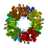
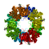
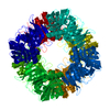
| Entry | Database: PDB / ID: 6l3w | ||||||
|---|---|---|---|---|---|---|---|
| Title | Crystal structure of BphC, a halotolerant catechol dioxygenase | ||||||
 Components Components | Extra-diol dioxygenase BphC | ||||||
 Keywords Keywords |  OXIDOREDUCTASE / Dioxygenase / catechol / metalloproteins / halotolerant OXIDOREDUCTASE / Dioxygenase / catechol / metalloproteins / halotolerant | ||||||
| Function / homology | 2,3-Dihydroxybiphenyl 1,2-Dioxygenase, domain 1 / 2,3-Dihydroxybiphenyl 1,2-Dioxygenase; domain 1 / Roll / Alpha Beta / :  Function and homology information Function and homology information | ||||||
| Biological species | metagenome (others) | ||||||
| Method | X-RAY DIFFRACTION / MOLECULAR REPLACEMENT / Resolution: 2.6 Å | ||||||
 Authors Authors | Thakur, K.G. / Solanki, V. | ||||||
| Funding support |  India, 1items India, 1items
| ||||||
 Citation Citation | Journal: mSystems / Year: 2019 Title: Structure Elucidation and Biochemical Characterization of Environmentally Relevant Novel Extradiol Dioxygenases Discovered by a Functional Metagenomics Approach. Authors: Sidhu, C. / Solanki, V. / Pinnaka, A.K. / Thakur, K.G. | ||||||
| History |
|
- Structure visualization
| Structure viewer | Molecule: MolmilJmol/JSmol |
|---|
- Downloads & links
-Download
| PDBx/mmCIF format | 6l3w.cif.gz | 78 KB | Display | PDBx/mmCIF format |
|---|---|---|---|---|
| PDB format | pdb6l3w.ent.gz | 55.4 KB | Display | PDB format |
| PDBx/mmJSON format | 6l3w.json.gz | Tree view | PDBx/mmJSON format | |
| Others |  Other downloads Other downloads |
-Validation report
| Arichive directory | https://data.pdbj.org/pub/pdb/validation_reports/l3/6l3wftp://data.pdbj.org/pub/pdb/validation_reports/l3/6l3w | HTTPS FTP |
|---|
-Related structure data
| Related structure data | 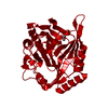 1hanS S: Starting model for refinement |
|---|---|
| Similar structure data |


-Links
 PDBj
PDBj- Assembly
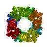
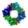
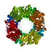
| Deposited unit | 
| ||||||||
|---|---|---|---|---|---|---|---|---|---|
| 1 | 
| ||||||||
| Unit cell |
| ||||||||
| Components on special symmetry positions |
|
-Components
| #1: Protein | Mass: 35253.914 Da / Num. of mol.: 1 Source method: isolated from a genetically manipulated source Source: (gene. exp.) metagenome (others) / Gene: 2,3-dihydroxybiphenyl 1,2-dioxygenase / Plasmid: pET28a / Cell (production host): BL21-DE3 / Production host:  Escherichia coli K-12 (bacteria) / Strain (production host): K-12 Escherichia coli K-12 (bacteria) / Strain (production host): K-12 |
|---|---|
| #2: Chemical | ChemComp-FE / Iron  Mass: 55.845 Da / Num. of mol.: 1 / Source method: obtained synthetically / Formula: Fe / Feature type: SUBJECT OF INVESTIGATION Mass: 55.845 Da / Num. of mol.: 1 / Source method: obtained synthetically / Formula: Fe / Feature type: SUBJECT OF INVESTIGATION |
| #3: Water | ChemComp-HOH / Water Mass: 18.015 Da / Num. of mol.: 151 / Source method: isolated from a natural source / Formula: H2O Mass: 18.015 Da / Num. of mol.: 151 / Source method: isolated from a natural source / Formula: H2O |
| Has ligand of interest | Y |
| Sequence details | AUTHORS STATE THAT THE NCBI ACCESSION NUMBER IS KX965751 FOR THIS SEQUENCE. |
-Experimental details
-Experiment
| Experiment | Method: X-RAY DIFFRACTION / Number of used crystals: 1 |
|---|
- Sample preparation
| Crystal | Density Matthews: 2.89 Å3/Da / Density % sol: 57.51 % |
|---|---|
| Crystal grow | Temperature: 291 K / Method: vapor diffusion, sitting drop / pH: 6 / Details: 0.1M sodium citrate pH 6.0, 2M NaCl |
-Data collection
| Diffraction | Mean temperature: 100 K / Serial crystal experiment: N | |||||||||||||||||||||||||||
|---|---|---|---|---|---|---|---|---|---|---|---|---|---|---|---|---|---|---|---|---|---|---|---|---|---|---|---|---|
| Diffraction source | Source: ROTATING ANODE / Type: RIGAKU MICROMAX-007 HF / Wavelength: 1.5418 Å | |||||||||||||||||||||||||||
| Detector | Type: MAR scanner 345 mm plate / Detector: IMAGE PLATE / Date: Oct 28, 2016 | |||||||||||||||||||||||||||
| Radiation | Protocol: SINGLE WAVELENGTH / Monochromatic (M) / Laue (L): M / Scattering type: x-ray | |||||||||||||||||||||||||||
| Radiation wavelength | Wavelength: 1.5418 Å / Relative weight: 1 | |||||||||||||||||||||||||||
| Reflection | Resolution: 2.6→31.19 Å / Num. obs: 12365 / % possible obs: 99.8 % / Redundancy: 5.3 % / CC1/2: 0.991 / Rmerge(I) obs: 0.141 / Rpim(I) all: 0.068 / Rrim(I) all: 0.157 / Rsym value: 0.141 / Net I/σ(I): 10.4 | |||||||||||||||||||||||||||
| Reflection shell | Diffraction-ID: 1
|
- Processing
| Software |
| ||||||||||||||||||||||||||||||||||||||||||||||||||||||||||||
|---|---|---|---|---|---|---|---|---|---|---|---|---|---|---|---|---|---|---|---|---|---|---|---|---|---|---|---|---|---|---|---|---|---|---|---|---|---|---|---|---|---|---|---|---|---|---|---|---|---|---|---|---|---|---|---|---|---|---|---|---|---|
| Refinement | Method to determine structure: MOLECULAR REPLACEMENT Starting model: 1HAN Resolution: 2.6→31.19 Å / Cor.coef. Fo:Fc: 0.929 / Cor.coef. Fo:Fc free: 0.884 / SU B: 10.355 / SU ML: 0.214 / SU R Cruickshank DPI: 0.6048 / Cross valid method: THROUGHOUT / σ(F): 0 / ESU R: 0.605 / ESU R Free: 0.302 Details: HYDROGENS HAVE BEEN ADDED IN THE RIDING POSITIONS U VALUES : REFINED INDIVIDUALLY
| ||||||||||||||||||||||||||||||||||||||||||||||||||||||||||||
| Solvent computation | Ion probe radii: 0.8 Å / Shrinkage radii: 0.8 Å / VDW probe radii: 1.2 Å | ||||||||||||||||||||||||||||||||||||||||||||||||||||||||||||
| Displacement parameters | Biso max: 74.22 Å2 / Biso mean: 23.993 Å2 / Biso min: 1.44 Å2
| ||||||||||||||||||||||||||||||||||||||||||||||||||||||||||||
| Refinement step | Cycle: final / Resolution: 2.6→31.19 Å
| ||||||||||||||||||||||||||||||||||||||||||||||||||||||||||||
| Refine LS restraints |
| ||||||||||||||||||||||||||||||||||||||||||||||||||||||||||||
| LS refinement shell | Resolution: 2.6→2.667 Å / Rfactor Rfree error: 0 / Total num. of bins used: 20
|
