 Movie
Movie Controller

[English] 日本語
 Yorodumi
- PDB-6kyl: Crystal Structure of Phosphatidic acid Transporter Ups1/Mdm35 in ... -
+ Open data

- Basic information
| Entry | Database: PDB / ID: 6kyl | |||||||||
|---|---|---|---|---|---|---|---|---|---|---|
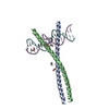
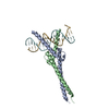
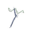
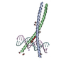
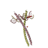
| Title | Crystal Structure of Phosphatidic acid Transporter Ups1/Mdm35 in Complex with (2R)-3-(phosphonooxy)propane-1,2-diyl dihexanoate | |||||||||
 Components Components |
| |||||||||
 Keywords Keywords |  TRANSPORT PROTEIN / Lipid transport TRANSPORT PROTEIN / Lipid transport | |||||||||
| Function / homology |  Function and homology information Function and homology informationTP53 Regulates Transcription of Genes Involved in Cytochrome C Release / phosphatidic acid transfer activity / cardiolipin metabolic process / positive regulation of phosphatidylcholine biosynthetic process / mitochondrial respiratory chain complex assembly / phospholipid transport / phospholipid translocation / mitochondrion organization / mitochondrial intermembrane space / mitochondrial inner membrane ...TP53 Regulates Transcription of Genes Involved in Cytochrome C Release / phosphatidic acid transfer activity / cardiolipin metabolic process / positive regulation of phosphatidylcholine biosynthetic process / mitochondrial respiratory chain complex assembly / phospholipid transport / phospholipid translocation / mitochondrion organization / mitochondrial intermembrane space / mitochondrial inner membrane / lipid binding / mitochondrion / nucleus / cytosol / cytoplasmSimilarity search - Function | |||||||||
| Biological species |  Saccharomyces cerevisiae S288c (yeast) Saccharomyces cerevisiae S288c (yeast) | |||||||||
| Method | X-RAY DIFFRACTION / SYNCHROTRON / MOLECULAR REPLACEMENT / Resolution: 3.55 Å | |||||||||
 Authors Authors | Lu, J. / Chan, K.C. / Zhai, Y. / Fan, J. / Sun, F. | |||||||||
 Citation Citation | Journal: Commun Biol / Year: 2020 Title: Molecular mechanism of mitochondrial phosphatidate transfer by Ups1. Authors: Lu, J. / Chan, C. / Yu, L. / Fan, J. / Sun, F. / Zhai, Y. | |||||||||
| History |
|
- Structure visualization
| Structure viewer | Molecule: MolmilJmol/JSmol |
|---|
- Downloads & links
-Download
| PDBx/mmCIF format | 6kyl.cif.gz | 180.3 KB | Display | PDBx/mmCIF format |
|---|---|---|---|---|
| PDB format | pdb6kyl.ent.gz | 140.8 KB | Display | PDB format |
| PDBx/mmJSON format | 6kyl.json.gz | Tree view | PDBx/mmJSON format | |
| Others |  Other downloads Other downloads |
-Validation report
| Arichive directory | https://data.pdbj.org/pub/pdb/validation_reports/ky/6kylftp://data.pdbj.org/pub/pdb/validation_reports/ky/6kyl | HTTPS FTP |
|---|
-Related structure data
| Related structure data | 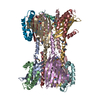 5jqlS S: Starting model for refinement |
|---|---|
| Similar structure data |


-Links
 PDBj
PDBj

- Assembly
| Deposited unit | 
| ||||||||
|---|---|---|---|---|---|---|---|---|---|
| 1 | 
| ||||||||
| 2 | 
| ||||||||
| Unit cell |
|
-Components
| #1: Protein | Mass: 9723.954 Da / Num. of mol.: 2 Source method: isolated from a genetically manipulated source Source: (gene. exp.) Saccharomyces cerevisiae S288c (yeast) / Gene: MDM35, YKL053C-A / Production host:  Escherichia coli (E. coli) / References: UniProt: O60200 Escherichia coli (E. coli) / References: UniProt: O60200#2: Protein | Mass: 21755.670 Da / Num. of mol.: 2 Source method: isolated from a genetically manipulated source Source: (gene. exp.) Saccharomyces cerevisiae S288c (yeast) / Gene: UPS1, YLR193C / Production host: Escherichia coli (E. coli) / References: UniProt: Q05776#3: Chemical |   Mass: 368.360 Da / Num. of mol.: 2 / Source method: obtained synthetically / Formula: C15H29O8P / Feature type: SUBJECT OF INVESTIGATION / Comment: phospholipid*YM Mass: 368.360 Da / Num. of mol.: 2 / Source method: obtained synthetically / Formula: C15H29O8P / Feature type: SUBJECT OF INVESTIGATION / Comment: phospholipid*YMHas ligand of interest | Y | |
|---|
-Experimental details
-Experiment
| Experiment | Method: X-RAY DIFFRACTION / Number of used crystals: 1 |
|---|
- Sample preparation
| Crystal | Density Matthews: 2.62 Å3/Da / Density % sol: 53.12 % |
|---|---|
| Crystal grow | Temperature: 289 K / Method: vapor diffusion, hanging drop Details: 0.1M Bis-tris Propane, 30%-32% v/v Tacsimate, pH7.0 |
-Data collection
| Diffraction | Mean temperature: 100 K / Serial crystal experiment: N | ||||||||||||||||||||||||||||||||||||||||||||||||||||||||||||||||||||||||||||||||||||||||||||||||||||||||||||||||||||||||||||||||||||||||||||||||||||||||||||||||||||||||
|---|---|---|---|---|---|---|---|---|---|---|---|---|---|---|---|---|---|---|---|---|---|---|---|---|---|---|---|---|---|---|---|---|---|---|---|---|---|---|---|---|---|---|---|---|---|---|---|---|---|---|---|---|---|---|---|---|---|---|---|---|---|---|---|---|---|---|---|---|---|---|---|---|---|---|---|---|---|---|---|---|---|---|---|---|---|---|---|---|---|---|---|---|---|---|---|---|---|---|---|---|---|---|---|---|---|---|---|---|---|---|---|---|---|---|---|---|---|---|---|---|---|---|---|---|---|---|---|---|---|---|---|---|---|---|---|---|---|---|---|---|---|---|---|---|---|---|---|---|---|---|---|---|---|---|---|---|---|---|---|---|---|---|---|---|---|---|---|---|---|
| Diffraction source | Source: SYNCHROTRON / Site: SSRF  / Beamline: BL18U1 / Wavelength: 0.9786 Å / Beamline: BL18U1 / Wavelength: 0.9786 Å | ||||||||||||||||||||||||||||||||||||||||||||||||||||||||||||||||||||||||||||||||||||||||||||||||||||||||||||||||||||||||||||||||||||||||||||||||||||||||||||||||||||||||
| Detector | Type: Bruker Platinum 135 / Detector: CCD / Date: Jul 6, 2015 | ||||||||||||||||||||||||||||||||||||||||||||||||||||||||||||||||||||||||||||||||||||||||||||||||||||||||||||||||||||||||||||||||||||||||||||||||||||||||||||||||||||||||
| Radiation | Protocol: SINGLE WAVELENGTH / Monochromatic (M) / Laue (L): M / Scattering type: x-ray | ||||||||||||||||||||||||||||||||||||||||||||||||||||||||||||||||||||||||||||||||||||||||||||||||||||||||||||||||||||||||||||||||||||||||||||||||||||||||||||||||||||||||
| Radiation wavelength | Wavelength: 0.9786 Å / Relative weight: 1 | ||||||||||||||||||||||||||||||||||||||||||||||||||||||||||||||||||||||||||||||||||||||||||||||||||||||||||||||||||||||||||||||||||||||||||||||||||||||||||||||||||||||||
| Reflection | Resolution: 3.549→50 Å / Num. obs: 8015 / % possible obs: 100 % / Redundancy: 6.7 % / Rmerge(I) obs: 0.053 / Rpim(I) all: 0.022 / Rrim(I) all: 0.057 / Χ2: 1 / Net I/σ(I): 6.4 | ||||||||||||||||||||||||||||||||||||||||||||||||||||||||||||||||||||||||||||||||||||||||||||||||||||||||||||||||||||||||||||||||||||||||||||||||||||||||||||||||||||||||
| Reflection shell | Diffraction-ID: 1 / % possible all: 100
|
- Processing
| Software |
| ||||||||||||||||||||||||||||||||||||||||||
|---|---|---|---|---|---|---|---|---|---|---|---|---|---|---|---|---|---|---|---|---|---|---|---|---|---|---|---|---|---|---|---|---|---|---|---|---|---|---|---|---|---|---|---|
| Refinement | Method to determine structure: MOLECULAR REPLACEMENT Starting model: 5JQL Resolution: 3.55→45.07 Å / SU ML: 0.52 / Cross valid method: THROUGHOUT / σ(F): 1.35 / Phase error: 33.11
| ||||||||||||||||||||||||||||||||||||||||||
| Solvent computation | Shrinkage radii: 0.9 Å / VDW probe radii: 1.11 Å | ||||||||||||||||||||||||||||||||||||||||||
| Displacement parameters | Biso max: 90.09 Å2 / Biso mean: 34.7814 Å2 / Biso min: 21.15 Å2 | ||||||||||||||||||||||||||||||||||||||||||
| Refinement step | Cycle: final / Resolution: 3.55→45.07 Å
| ||||||||||||||||||||||||||||||||||||||||||
| LS refinement shell | Refine-ID: X-RAY DIFFRACTION / Rfactor Rfree error: 0
| ||||||||||||||||||||||||||||||||||||||||||
| Refinement TLS params. | Method: refined / Origin x: -0.6368 Å / Origin y: 45.9889 Å / Origin z: -9.4485 Å
| ||||||||||||||||||||||||||||||||||||||||||
| Refinement TLS group |
|
