 Movie
Movie Controller


+ Open data

- Basic information
| Entry | Database: PDB / ID: 6kxt | ||||||
|---|---|---|---|---|---|---|---|
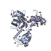
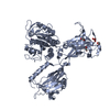
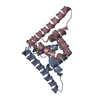
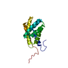
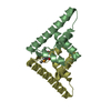
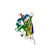
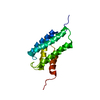
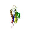
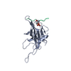
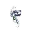
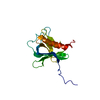
| Title | BON1-C2B | ||||||
 Components Components | Protein BONZAI 1 | ||||||
 Keywords Keywords | LIPID BINDING PROTEIN /  Copines / BON1 / C2 domain / Phospholipid / Membrane. Copines / BON1 / C2 domain / Phospholipid / Membrane. | ||||||
| Function / homology |  Function and homology information Function and homology informationresponse to humidity / plasmodesma / calcium-dependent phospholipid binding / plastid / response to temperature stimulus / positive regulation of cell size / defense response / metal ion binding / plasma membraneSimilarity search - Function | ||||||
| Biological species |  Arabidopsis thaliana (thale cress) Arabidopsis thaliana (thale cress) | ||||||
| Method | X-RAY DIFFRACTION / SYNCHROTRON / MOLECULAR REPLACEMENT / Resolution: 1.25 Å | ||||||
 Authors Authors | Wang, Q.C. / Jiang, M.Q. / Isupov, M.N. / Sun, L.F. / Wu, Y.K. | ||||||
 Citation Citation | Journal: To be published Title: Crystal Structure of an Arabidopsis Copine providing insights into this protein family Authors: Wang, Q.C. / Jiang, M.Q. / Isupov, M.N. / Sun, L.F. / Wu, Y.K. | ||||||
| History |
|
- Structure visualization
| Structure viewer | Molecule: MolmilJmol/JSmol |
|---|
- Downloads & links
-Download
| PDBx/mmCIF format | 6kxt.cif.gz | 49.6 KB | Display | PDBx/mmCIF format |
|---|---|---|---|---|
| PDB format | pdb6kxt.ent.gz | 33.9 KB | Display | PDB format |
| PDBx/mmJSON format | 6kxt.json.gz | Tree view | PDBx/mmJSON format | |
| Others |  Other downloads Other downloads |
-Validation report
| Arichive directory | https://data.pdbj.org/pub/pdb/validation_reports/kx/6kxtftp://data.pdbj.org/pub/pdb/validation_reports/kx/6kxt | HTTPS FTP |
|---|
-Related structure data

-Links
 PDBj
PDBj


- Assembly
| Deposited unit | 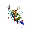
| |||||||||||||||
|---|---|---|---|---|---|---|---|---|---|---|---|---|---|---|---|---|
| 1 |
| |||||||||||||||
| Unit cell |
| |||||||||||||||
| Components on special symmetry positions |
|
-Components
| #1: Protein | Mass: 15680.918 Da / Num. of mol.: 1 Source method: isolated from a genetically manipulated source Source: (gene. exp.) Arabidopsis thaliana (thale cress) / Gene: BON1, CPN1, At5g61900, K22G18.2 / Production host:  Escherichia coli BL21(DE3) (bacteria) / References: UniProt: Q941L3 Escherichia coli BL21(DE3) (bacteria) / References: UniProt: Q941L3 | ||||
|---|---|---|---|---|---|
| #2: Chemical | ChemComp-MES / MES (buffer)  Mass: 195.237 Da / Num. of mol.: 1 / Source method: obtained synthetically / Formula: C6H13NO4S / Comment: pH buffer*YM Mass: 195.237 Da / Num. of mol.: 1 / Source method: obtained synthetically / Formula: C6H13NO4S / Comment: pH buffer*YM | ||||
| #3: Chemical | Sulfate  Mass: 96.063 Da / Num. of mol.: 2 / Source method: obtained synthetically / Formula: SO4 Mass: 96.063 Da / Num. of mol.: 2 / Source method: obtained synthetically / Formula: SO4#4: Water | ChemComp-HOH / | Water Mass: 18.015 Da / Num. of mol.: 200 / Source method: isolated from a natural source / Formula: H2O Mass: 18.015 Da / Num. of mol.: 200 / Source method: isolated from a natural source / Formula: H2OHas ligand of interest | N | |
-Experimental details
-Experiment
| Experiment | Method: X-RAY DIFFRACTION / Number of used crystals: 1 |
|---|
- Sample preparation
| Crystal | Density Matthews: 2.05 Å3/Da / Density % sol: 40.12 % |
|---|---|
| Crystal grow | Temperature: 289 K / Method: vapor diffusion, sitting drop / Details: Two moles of ammonium sulfate |
-Data collection
| Diffraction | Mean temperature: 100 K / Serial crystal experiment: N | ||||||||||||||||||||||||
|---|---|---|---|---|---|---|---|---|---|---|---|---|---|---|---|---|---|---|---|---|---|---|---|---|---|
| Diffraction source | Source: SYNCHROTRON / Site: SSRF  / Beamline: BL19U1 / Wavelength: 0.9789 Å / Beamline: BL19U1 / Wavelength: 0.9789 Å | ||||||||||||||||||||||||
| Detector | Type: DECTRIS PILATUS 6M / Detector: PIXEL / Date: Sep 10, 2018 | ||||||||||||||||||||||||
| Radiation | Protocol: SINGLE WAVELENGTH / Monochromatic (M) / Laue (L): M / Scattering type: x-ray | ||||||||||||||||||||||||
| Radiation wavelength | Wavelength: 0.9789 Å / Relative weight: 1 | ||||||||||||||||||||||||
| Reflection | Resolution: 1.25→40.73 Å / Num. obs: 32577 / % possible obs: 90.1 % / Redundancy: 12.2 % / CC1/2: 1 / Rmerge(I) obs: 0.073 / Rpim(I) all: 0.021 / Rrim(I) all: 0.076 / Net I/σ(I): 16.4 | ||||||||||||||||||||||||
| Reflection shell | Diffraction-ID: 1
|
- Processing
| Software |
| ||||||||||||||||||||||||||||||||||||||||||||||||||||||||||||
|---|---|---|---|---|---|---|---|---|---|---|---|---|---|---|---|---|---|---|---|---|---|---|---|---|---|---|---|---|---|---|---|---|---|---|---|---|---|---|---|---|---|---|---|---|---|---|---|---|---|---|---|---|---|---|---|---|---|---|---|---|---|
| Refinement | Method to determine structure: MOLECULAR REPLACEMENT / Resolution: 1.25→40.73 Å / Cor.coef. Fo:Fc: 0.977 / Cor.coef. Fo:Fc free: 0.974 / SU B: 1.284 / SU ML: 0.05 / Cross valid method: THROUGHOUT / σ(F): 0 / ESU R: 0.053 / ESU R Free: 0.055 Details: HYDROGENS HAVE BEEN ADDED IN THE RIDING POSITIONS U VALUES : REFINED INDIVIDUALLY
| ||||||||||||||||||||||||||||||||||||||||||||||||||||||||||||
| Solvent computation | Ion probe radii: 0.8 Å / Shrinkage radii: 0.8 Å / VDW probe radii: 1.2 Å | ||||||||||||||||||||||||||||||||||||||||||||||||||||||||||||
| Displacement parameters | Biso max: 75.67 Å2 / Biso mean: 20.071 Å2 / Biso min: 11.57 Å2
| ||||||||||||||||||||||||||||||||||||||||||||||||||||||||||||
| Refinement step | Cycle: final / Resolution: 1.25→40.73 Å
| ||||||||||||||||||||||||||||||||||||||||||||||||||||||||||||
| Refine LS restraints |
| ||||||||||||||||||||||||||||||||||||||||||||||||||||||||||||
| LS refinement shell | Resolution: 1.25→1.282 Å / Rfactor Rfree error: 0
|
