 Movie
Movie Controller


+ Open data

- Basic information
| Entry | Database: PDB / ID: 6kxb | ||||||
|---|---|---|---|---|---|---|---|
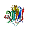
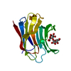
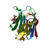
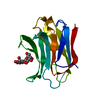
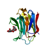
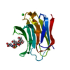
| Title | Galectin-3 CRD binds to GalA trimer | ||||||
 Components Components | Galectin-3 | ||||||
 Keywords Keywords | SUGAR BINDING PROTEIN / Galectin-3 CRD / GalA trimer | ||||||
| Function / homology |  Function and homology information Function and homology informationnegative regulation of protein tyrosine phosphatase activity / negative regulation of immunological synapse formation / RUNX2 regulates genes involved in differentiation of myeloid cells / negative regulation of T cell activation via T cell receptor contact with antigen bound to MHC molecule on antigen presenting cell / regulation of T cell apoptotic process / mononuclear cell migration / IgE binding / positive regulation of mononuclear cell migration / negative regulation of endocytosis / eosinophil chemotaxis ...negative regulation of protein tyrosine phosphatase activity / negative regulation of immunological synapse formation / RUNX2 regulates genes involved in differentiation of myeloid cells / negative regulation of T cell activation via T cell receptor contact with antigen bound to MHC molecule on antigen presenting cell / regulation of T cell apoptotic process / mononuclear cell migration / IgE binding / positive regulation of mononuclear cell migration / negative regulation of endocytosis / eosinophil chemotaxis / regulation of extrinsic apoptotic signaling pathway via death domain receptors / RUNX1 regulates transcription of genes involved in differentiation of myeloid cells / protein phosphatase inhibitor activity / negative regulation of T cell receptor signaling pathway / macrophage chemotaxis / positive chemotaxis / regulation of T cell proliferation / positive regulation of calcium ion import / chemoattractant activity / monocyte chemotaxis / Advanced glycosylation endproduct receptor signaling / ficolin-1-rich granule membrane / immunological synapse / laminin binding / epithelial cell differentiation / molecular condensate scaffold activity / neutrophil chemotaxis / secretory granule membrane / RNA splicing / negative regulation of extrinsic apoptotic signaling pathway / positive regulation of protein-containing complex assembly / positive regulation of protein localization to plasma membrane / spliceosomal complex / mRNA processing / carbohydrate binding / protein phosphatase binding / collagen-containing extracellular matrix / mitochondrial inner membrane / innate immune response / Neutrophil degranulation / cell surface / extracellular space / RNA binding / extracellular exosome / extracellular region / nucleoplasm / membrane / nucleus / plasma membrane / cytosol / cytoplasmSimilarity search - Function | ||||||
| Biological species |  Homo sapiens (human) Homo sapiens (human) | ||||||
| Method | X-RAY DIFFRACTION / SYNCHROTRON / MOLECULAR REPLACEMENT / Resolution: 1.5 Å | ||||||
 Authors Authors | Su, J. | ||||||
 Citation Citation | Journal: Glycobiology / Year: 2021 Title: Topsy-turvy binding of negatively charged homogalacturonan oligosaccharides to galectin-3. Authors: Zheng, Y. / Su, J. / Miller, M.C. / Geng, J. / Xu, X. / Zhang, T. / Mayzel, M. / Zhou, Y. / Mayo, K.H. / Tai, G. | ||||||
| History |
|
- Structure visualization
| Structure viewer | Molecule: MolmilJmol/JSmol |
|---|
- Downloads & links
-Download
| PDBx/mmCIF format | 6kxb.cif.gz | 102.3 KB | Display | PDBx/mmCIF format |
|---|---|---|---|---|
| PDB format | pdb6kxb.ent.gz | 77.9 KB | Display | PDB format |
| PDBx/mmJSON format | 6kxb.json.gz | Tree view | PDBx/mmJSON format | |
| Others |  Other downloads Other downloads |
-Validation report
| Arichive directory | https://data.pdbj.org/pub/pdb/validation_reports/kx/6kxbftp://data.pdbj.org/pub/pdb/validation_reports/kx/6kxb | HTTPS FTP |
|---|
-Related structure data
| Related structure data |  6kxaC  4r9aS S: Starting model for refinement C: citing same article ( |
|---|---|
| Similar structure data |


-Links
 PDBj
PDBj


- Assembly
| Deposited unit | 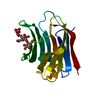
| ||||||||
|---|---|---|---|---|---|---|---|---|---|
| 1 |
| ||||||||
| Unit cell |
|
-Components
| #1: Protein | / Gal-3 / 35 kDa lectin / Carbohydrate-binding protein 35 / CBP 35 / Galactose-specific lectin 3 / ...Gal-3 / 35 kDa lectin / Carbohydrate-binding protein 35 / CBP 35 / Galactose-specific lectin 3 / Galactoside-binding protein / GALBP / IgE-binding protein / L-31 / Laminin-binding protein / Lectin L-29 / Mac-2 antigen Mass: 15701.049 Da / Num. of mol.: 1 Source method: isolated from a genetically manipulated source Source: (gene. exp.) Homo sapiens (human) / Gene: LGALS3, MAC2 / Production host:  Escherichia coli (E. coli) / References: UniProt: P17931 Escherichia coli (E. coli) / References: UniProt: P17931 |
|---|---|
| #2: Polysaccharide | alpha-D-galactopyranuronic acid-(1-4)-alpha-D-galactopyranuronic acid-(1-4)-beta-D-galactopyranuronic acid / Mass: 546.387 Da / Num. of mol.: 1 Source method: isolated from a genetically manipulated source |
| #3: Water | ChemComp-HOH / Water Mass: 18.015 Da / Num. of mol.: 201 / Source method: isolated from a natural source / Formula: H2O Mass: 18.015 Da / Num. of mol.: 201 / Source method: isolated from a natural source / Formula: H2O |
| Has ligand of interest | Y |
-Experimental details
-Experiment
| Experiment | Method: X-RAY DIFFRACTION / Number of used crystals: 1 |
|---|
- Sample preparation
| Crystal | Density Matthews: 2.03 Å3/Da / Density % sol: 39.55 % |
|---|---|
| Crystal grow | Temperature: 298 K / Method: vapor diffusion, hanging drop / Details: PEG |
-Data collection
| Diffraction | Mean temperature: 100 K / Serial crystal experiment: N |
|---|---|
| Diffraction source | Source: SYNCHROTRON / Site: SSRF  / Beamline: BL18U1 / Wavelength: 0.97 Å / Beamline: BL18U1 / Wavelength: 0.97 Å |
| Detector | Type: DECTRIS PILATUS3 6M / Detector: PIXEL / Date: Jun 19, 2018 |
| Radiation | Protocol: SINGLE WAVELENGTH / Monochromatic (M) / Laue (L): M / Scattering type: x-ray |
| Radiation wavelength | Wavelength: 0.97 Å / Relative weight: 1 |
| Reflection | Resolution: 1.5→19.34 Å / Num. obs: 21105 / % possible obs: 99.7 % / Redundancy: 5.8 % / Rmerge(I) obs: 0.07 / Net I/σ(I): 15.4 |
| Reflection shell | Resolution: 1.5→1.53 Å / Rmerge(I) obs: 0.195 |
- Processing
| Software |
| |||||||||||||||||||||||||||||||||||||||||||||||||||||||||||||||||||||||||||||||||||||||||||||||||||||||||
|---|---|---|---|---|---|---|---|---|---|---|---|---|---|---|---|---|---|---|---|---|---|---|---|---|---|---|---|---|---|---|---|---|---|---|---|---|---|---|---|---|---|---|---|---|---|---|---|---|---|---|---|---|---|---|---|---|---|---|---|---|---|---|---|---|---|---|---|---|---|---|---|---|---|---|---|---|---|---|---|---|---|---|---|---|---|---|---|---|---|---|---|---|---|---|---|---|---|---|---|---|---|---|---|---|---|---|
| Refinement | Method to determine structure: MOLECULAR REPLACEMENT Starting model: 4R9A Resolution: 1.5→19.34 Å / SU ML: 0.15 / σ(F): 1.4 / Phase error: 19.72 / Stereochemistry target values: ML
| |||||||||||||||||||||||||||||||||||||||||||||||||||||||||||||||||||||||||||||||||||||||||||||||||||||||||
| Solvent computation | Shrinkage radii: 0.9 Å / VDW probe radii: 1.11 Å / Solvent model: FLAT BULK SOLVENT MODEL | |||||||||||||||||||||||||||||||||||||||||||||||||||||||||||||||||||||||||||||||||||||||||||||||||||||||||
| Displacement parameters | Biso mean: 21.99 Å2 | |||||||||||||||||||||||||||||||||||||||||||||||||||||||||||||||||||||||||||||||||||||||||||||||||||||||||
| Refinement step | Cycle: LAST / Resolution: 1.5→19.34 Å
| |||||||||||||||||||||||||||||||||||||||||||||||||||||||||||||||||||||||||||||||||||||||||||||||||||||||||
| LS refinement shell |
|
