 Movie
Movie Controller

[English] 日本語
 Yorodumi
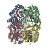
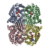
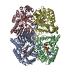
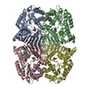
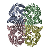
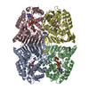
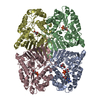
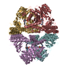
- PDB-6ktl: Crystal structure of scyllo-inositol dehydrogenase R178A mutant, ... -
+ Open data

- Basic information
| Entry | Database: PDB / ID: 6ktl | ||||||
|---|---|---|---|---|---|---|---|
| Title | Crystal structure of scyllo-inositol dehydrogenase R178A mutant, complexed with NAD and myo-inositol, from Paracoccus laeviglucosivorans | ||||||
 Components Components | Scyllo-inositol dehydrogenase with L-glucose dehydrogenase activity | ||||||
 Keywords Keywords |  OXIDOREDUCTASE OXIDOREDUCTASE | ||||||
| Function / homology |  Function and homology information Function and homology information | ||||||
| Biological species |  Paracoccus laeviglucosivorans (bacteria) Paracoccus laeviglucosivorans (bacteria) | ||||||
| Method | X-RAY DIFFRACTION / SYNCHROTRON / MOLECULAR REPLACEMENT / Resolution: 1.65 Å | ||||||
 Authors Authors | Suzuki, M. / Koubara, K. / Takenoya, M. / Fukano, K. / Ito, S. / Sasaki, Y. / Nakamura, A. / Yajima, S. | ||||||
| Funding support |  Japan, 1items Japan, 1items
| ||||||
 Citation Citation | Journal: Biosci.Biotechnol.Biochem. / Year: 2020 Title: Single amino acid mutation altered substrate specificity for L-glucose and inositol inscyllo-inositol dehydrogenase isolated fromParacoccus laeviglucosivorans. Authors: Suzuki, M. / Koubara, K. / Takenoya, M. / Fukano, K. / Ito, S. / Sasaki, Y. / Nakamura, A. / Yajima, S. | ||||||
| History |
|
- Structure visualization
| Structure viewer | Molecule: MolmilJmol/JSmol |
|---|
- Downloads & links
-Download
| PDBx/mmCIF format | 6ktl.cif.gz | 314.5 KB | Display | PDBx/mmCIF format |
|---|---|---|---|---|
| PDB format | pdb6ktl.ent.gz | 252.3 KB | Display | PDB format |
| PDBx/mmJSON format | 6ktl.json.gz | Tree view | PDBx/mmJSON format | |
| Others |  Other downloads Other downloads |
-Validation report
| Arichive directory | https://data.pdbj.org/pub/pdb/validation_reports/kt/6ktlftp://data.pdbj.org/pub/pdb/validation_reports/kt/6ktl | HTTPS FTP |
|---|
-Related structure data
| Related structure data |  6ktjC  6ktkC  5yabS S: Starting model for refinement C: citing same article ( |
|---|---|
| Similar structure data |


-Links
 PDBj
PDBj- Assembly
| Deposited unit | 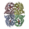
| ||||||||
|---|---|---|---|---|---|---|---|---|---|
| 1 |
| ||||||||
| Unit cell |
|
-Components
| #1: Protein | Mass: 41315.594 Da / Num. of mol.: 4 / Mutation: R178A Source method: isolated from a genetically manipulated source Source: (gene. exp.) Paracoccus laeviglucosivorans (bacteria)Gene: lgdA / Production host: Escherichia coli BL21(DE3) (bacteria) / References: UniProt: K7ZP76#2: Chemical | Acetate  Mass: 59.044 Da / Num. of mol.: 3 / Source method: obtained synthetically / Formula: C2H3O2 Mass: 59.044 Da / Num. of mol.: 3 / Source method: obtained synthetically / Formula: C2H3O2#3: Chemical | ChemComp-NAD / Nicotinamide adenine dinucleotide  Mass: 663.425 Da / Num. of mol.: 4 / Source method: obtained synthetically / Formula: C21H27N7O14P2 / Comment: NAD*YM Mass: 663.425 Da / Num. of mol.: 4 / Source method: obtained synthetically / Formula: C21H27N7O14P2 / Comment: NAD*YM#4: Chemical | ChemComp-INS / | Inositol  Mass: 180.156 Da / Num. of mol.: 1 / Source method: obtained synthetically / Formula: C6H12O6 / Feature type: SUBJECT OF INVESTIGATION / Comment: neurotransmitter, hormone*YM Mass: 180.156 Da / Num. of mol.: 1 / Source method: obtained synthetically / Formula: C6H12O6 / Feature type: SUBJECT OF INVESTIGATION / Comment: neurotransmitter, hormone*YM#5: Water | ChemComp-HOH / | Water Mass: 18.015 Da / Num. of mol.: 1171 / Source method: isolated from a natural source / Formula: H2O Mass: 18.015 Da / Num. of mol.: 1171 / Source method: isolated from a natural source / Formula: H2OHas ligand of interest | Y | Sequence details | unintended mutation | |
|---|
-Experimental details
-Experiment
| Experiment | Method: X-RAY DIFFRACTION / Number of used crystals: 1 |
|---|
- Sample preparation
| Crystal | Density Matthews: 2.51 Å3/Da / Density % sol: 51 % |
|---|---|
| Crystal grow | Temperature: 293 K / Method: vapor diffusion, hanging drop / Details: 0.1 M sodium citrate, pH 5.2, and 20% PEG3350 |
-Data collection
| Diffraction | Mean temperature: 95 K / Serial crystal experiment: N |
|---|---|
| Diffraction source | Source: SYNCHROTRON / Site: Photon Factory / Beamline: BL-17A / Wavelength: 0.98 Å |
| Detector | Type: DECTRIS EIGER X 16M / Detector: PIXEL / Date: Dec 25, 2018 |
| Radiation | Protocol: SINGLE WAVELENGTH / Monochromatic (M) / Laue (L): M / Scattering type: x-ray |
| Radiation wavelength | Wavelength: 0.98 Å / Relative weight: 1 |
| Reflection | Resolution: 1.65→50 Å / Num. obs: 191340 / % possible obs: 99.6 % / Redundancy: 6.6 % / CC1/2: 0.999 / Rmerge(I) obs: 0.073 / Net I/σ(I): 16 |
| Reflection shell | Resolution: 1.65→1.69 Å / Rmerge(I) obs: 0.55 / Mean I/σ(I) obs: 4.4 / Num. unique obs: 13572 / CC1/2: 0.784 |
- Processing
| Software |
| ||||||||||||||||
|---|---|---|---|---|---|---|---|---|---|---|---|---|---|---|---|---|---|
| Refinement | Method to determine structure: MOLECULAR REPLACEMENT Starting model: 5yab Resolution: 1.65→48.9 Å / Cross valid method: THROUGHOUT
| ||||||||||||||||
| Refinement step | Cycle: LAST / Resolution: 1.65→48.9 Å
| ||||||||||||||||
| LS refinement shell | Resolution: 1.65→1.69 Å
|
