 Movie
Movie Controller


+ Open data

- Basic information
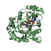
| Entry | Database: PDB / ID: 6ksg | ||||||
|---|---|---|---|---|---|---|---|
| Title | Vibrio cholerae Methionine Aminopeptidase in holo form | ||||||
 Components Components | Methionine aminopeptidase Methionyl aminopeptidase Methionyl aminopeptidase | ||||||
 Keywords Keywords | HYDROLASE | ||||||
| Function / homology |  Function and homology information Function and homology informationinitiator methionyl aminopeptidase activity / methionyl aminopeptidase / metalloaminopeptidase activity / transition metal ion binding / proteolysis / cytosolSimilarity search - Function | ||||||
| Biological species |  Vibrio cholerae serotype O1 (bacteria) Vibrio cholerae serotype O1 (bacteria) | ||||||
| Method | X-RAY DIFFRACTION / SYNCHROTRON / MOLECULAR REPLACEMENT / molecular replacement / Resolution: 1.9 Å | ||||||
 Authors Authors | Pillalamarri, V. / Addlagatta, A. | ||||||
| Funding support |  India, 1items India, 1items
| ||||||
 Citation Citation | Journal: Eur.J.Med.Chem. / Year: 2020 Title: Methionine aminopeptidases with short sequence inserts within the catalytic domain are differentially inhibited: Structural and biochemical studies of three proteins from Vibrio spp. Authors: Pillalamarri, V. / Reddy, C.G. / Bala, S.C. / Jangam, A. / Kutty, V.V. / Addlagatta, A. | ||||||
| History |
|
- Structure visualization
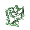
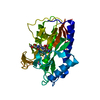
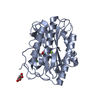
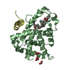
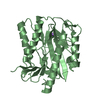
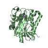
| Structure viewer | Molecule: MolmilJmol/JSmol |
|---|
- Downloads & links
-Download
| PDBx/mmCIF format | 6ksg.cif.gz | 132.1 KB | Display | PDBx/mmCIF format |
|---|---|---|---|---|
| PDB format | pdb6ksg.ent.gz | 100.1 KB | Display | PDB format |
| PDBx/mmJSON format | 6ksg.json.gz | Tree view | PDBx/mmJSON format | |
| Others |  Other downloads Other downloads |
-Validation report
| Arichive directory | https://data.pdbj.org/pub/pdb/validation_reports/ks/6ksgftp://data.pdbj.org/pub/pdb/validation_reports/ks/6ksg | HTTPS FTP |
|---|
-Related structure data
| Related structure data | 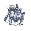 6k26SC  6lh7C S: Starting model for refinement C: citing same article ( |
|---|---|
| Similar structure data |


-Links
 PDBj
PDBj- Assembly
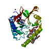
| Deposited unit | 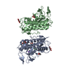
| ||||||||
|---|---|---|---|---|---|---|---|---|---|
| 1 | 
| ||||||||
| 2 | 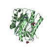
| ||||||||
| Unit cell |
|
-Components
| #1: Protein | Methionyl aminopeptidase / MetAP / Peptidase M Mass: 33573.352 Da / Num. of mol.: 2 Source method: isolated from a genetically manipulated source Source: (gene. exp.) Vibrio cholerae serotype O1 (strain ATCC 39315 / El Tor Inaba N16961) (bacteria)Strain: ATCC 39315 / El Tor Inaba N16961 / Gene: map, VC_2261 / Plasmid: pET15b / Production host: Escherichia coli BL21(DE3) (bacteria) / Strain (production host): BL21(DE3) / References: UniProt: Q9KPV1, methionyl aminopeptidase#2: Chemical | ChemComp-NI / Nickel  Mass: 58.693 Da / Num. of mol.: 4 / Source method: obtained synthetically / Formula: Ni Mass: 58.693 Da / Num. of mol.: 4 / Source method: obtained synthetically / Formula: Ni#3: Chemical |   Mass: 22.990 Da / Num. of mol.: 2 / Source method: obtained synthetically / Formula: Na Mass: 22.990 Da / Num. of mol.: 2 / Source method: obtained synthetically / Formula: Na#4: Chemical | ChemComp-GOL / Glycerol  Mass: 92.094 Da / Num. of mol.: 6 / Source method: obtained synthetically / Formula: C3H8O3 Mass: 92.094 Da / Num. of mol.: 6 / Source method: obtained synthetically / Formula: C3H8O3#5: Water | ChemComp-HOH / | Water Mass: 18.015 Da / Num. of mol.: 209 / Source method: isolated from a natural source / Formula: H2O Mass: 18.015 Da / Num. of mol.: 209 / Source method: isolated from a natural source / Formula: H2OHas ligand of interest | N | |
|---|
-Experimental details
-Experiment
| Experiment | Method: X-RAY DIFFRACTION / Number of used crystals: 1 |
|---|
- Sample preparation
| Crystal | Density Matthews: 2.56 Å3/Da / Density % sol: 51.96 % / Mosaicity: 0.24 ° |
|---|---|
| Crystal grow | Temperature: 298 K / Method: vapor diffusion, hanging drop / pH: 7.5 / Details: PEG 3350 |
-Data collection
| Diffraction | Mean temperature: 100 K / Serial crystal experiment: N | ||||||||||||||||||||||||||||||
|---|---|---|---|---|---|---|---|---|---|---|---|---|---|---|---|---|---|---|---|---|---|---|---|---|---|---|---|---|---|---|---|
| Diffraction source | Source: SYNCHROTRON / Site: ELETTRA  / Beamline: 11.2C / Wavelength: 0.9536 Å / Beamline: 11.2C / Wavelength: 0.9536 Å | ||||||||||||||||||||||||||||||
| Detector | Type: DECTRIS PILATUS 6M / Detector: PIXEL / Date: Jul 28, 2019 | ||||||||||||||||||||||||||||||
| Radiation | Protocol: SINGLE WAVELENGTH / Monochromatic (M) / Laue (L): M / Scattering type: x-ray | ||||||||||||||||||||||||||||||
| Radiation wavelength | Wavelength: 0.9536 Å / Relative weight: 1 | ||||||||||||||||||||||||||||||
| Reflection | Resolution: 1.9→48 Å / Num. obs: 50007 / % possible obs: 99.6 % / Redundancy: 6.1 % / CC1/2: 0.999 / Rmerge(I) obs: 0.074 / Rpim(I) all: 0.032 / Rrim(I) all: 0.081 / Net I/σ(I): 15.8 / Num. measured all: 307534 / Scaling rejects: 125 | ||||||||||||||||||||||||||||||
| Reflection shell | Diffraction-ID: 1
|
-Phasing
| Phasing | Method: molecular replacement | ||||||
|---|---|---|---|---|---|---|---|
| Phasing MR | R rigid body: 0.487
|
- Processing
| Software |
| ||||||||||||||||||||||||||||||||||||||||||||||||||||||||||||
|---|---|---|---|---|---|---|---|---|---|---|---|---|---|---|---|---|---|---|---|---|---|---|---|---|---|---|---|---|---|---|---|---|---|---|---|---|---|---|---|---|---|---|---|---|---|---|---|---|---|---|---|---|---|---|---|---|---|---|---|---|---|
| Refinement | Method to determine structure: MOLECULAR REPLACEMENT Starting model: 6K26 Resolution: 1.9→46.58 Å / Cor.coef. Fo:Fc: 0.946 / Cor.coef. Fo:Fc free: 0.916 / WRfactor Rfree: 0.2276 / WRfactor Rwork: 0.1867 / FOM work R set: 0.8286 / SU B: 3.566 / SU ML: 0.104 / SU R Cruickshank DPI: 0.153 / SU Rfree: 0.1456 / Cross valid method: THROUGHOUT / σ(F): 0 / ESU R: 0.153 / ESU R Free: 0.146 / Stereochemistry target values: MAXIMUM LIKELIHOOD / Details: MOLECULAR REPLCEMENT
| ||||||||||||||||||||||||||||||||||||||||||||||||||||||||||||
| Solvent computation | Ion probe radii: 0.8 Å / Shrinkage radii: 0.8 Å / VDW probe radii: 1.2 Å / Solvent model: MASK | ||||||||||||||||||||||||||||||||||||||||||||||||||||||||||||
| Displacement parameters | Biso max: 72.75 Å2 / Biso mean: 28.019 Å2 / Biso min: 8.36 Å2
| ||||||||||||||||||||||||||||||||||||||||||||||||||||||||||||
| Refinement step | Cycle: final / Resolution: 1.9→46.58 Å
| ||||||||||||||||||||||||||||||||||||||||||||||||||||||||||||
| Refine LS restraints |
| ||||||||||||||||||||||||||||||||||||||||||||||||||||||||||||
| LS refinement shell | Resolution: 1.903→1.952 Å / Rfactor Rfree error: 0 / Total num. of bins used: 20
|
