 Movie
Movie Controller


+ Open data

- Basic information
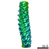
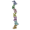
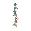
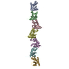
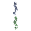
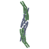
| Entry | Database: PDB / ID: 6kmf | ||||||||||||||||||
|---|---|---|---|---|---|---|---|---|---|---|---|---|---|---|---|---|---|---|---|
| Title | FimA type V pilus from P.gingivalis | ||||||||||||||||||
 Components Components | Major fimbrium subunit FimA type-1 | ||||||||||||||||||
 Keywords Keywords |  CELL ADHESION / Type V pilus / FimA / Porphyromonas gingivalis / ATCC33277 CELL ADHESION / Type V pilus / FimA / Porphyromonas gingivalis / ATCC33277 | ||||||||||||||||||
| Function / homology |  Function and homology informationpilus / cell outer membrane / cell adhesion / structural molecule activity Function and homology informationpilus / cell outer membrane / cell adhesion / structural molecule activitySimilarity search - Function | ||||||||||||||||||
| Biological species |  Porphyromonas gingivalis ATCC 33277 (bacteria) Porphyromonas gingivalis ATCC 33277 (bacteria) | ||||||||||||||||||
| Method | ELECTRON MICROSCOPY / single particle reconstruction / cryo EM / Resolution: 3.6 Å | ||||||||||||||||||
 Authors Authors | Shibata, S. / Shoji, M. / Matsunami, H. / Matthews, M. / Imada, K. / Nakayama, K. / Wolf, M. | ||||||||||||||||||
| Funding support |  Japan, 5items Japan, 5items
| ||||||||||||||||||
 Citation Citation | Journal: Nat Microbiol / Year: 2020 Title: Structure of polymerized type V pilin reveals assembly mechanism involving protease-mediated strand exchange. Authors: Satoshi Shibata / Mikio Shoji / Kodai Okada / Hideyuki Matsunami / Melissa M Matthews / Katsumi Imada / Koji Nakayama / Matthias Wolf / Abstract: Bacterial adhesion is a general strategy for host-microbe and microbe-microbe interactions. Adhesive pili are essential for colonization, biofilm formation, virulence and pathogenesis of many ...Bacterial adhesion is a general strategy for host-microbe and microbe-microbe interactions. Adhesive pili are essential for colonization, biofilm formation, virulence and pathogenesis of many environmental and pathogenic bacteria. Members of the class Bacteroidia have unique type V pili, assembled by protease-mediated polymerization. Porphyromonas gingivalis is the main contributor to periodontal disease and its type V pili are a key factor for its virulence. However, the structure of the polymerized pilus and its assembly mechanism are unknown. Here we show structures of polymerized and monomeric states of FimA stalk pilin from P. gingivalis, determined by cryo-electron microscopy and crystallography. The atomic model of assembled FimA shows that the C-terminal strand of a donor subunit is inserted into a groove in the β-sheet of an acceptor subunit after N-terminal cleavage by the protease RgpB. The C terminus of the donor strand is essential for polymerization. We propose that type V pili assemble via a sequential polar assembly mechanism at the cell surface, involving protease-mediated strand exchange, employed by various Gram-negative species belonging to the class Bacteroidia. Our results reveal functional surfaces related to pathogenic properties of polymerized FimA. These insights may facilitate development of antibacterial drugs. | ||||||||||||||||||
| History |
|
- Structure visualization
| Movie |
Movie viewer |
|---|---|
| Structure viewer | Molecule: MolmilJmol/JSmol |
- Downloads & links
-Download
| PDBx/mmCIF format | 6kmf.cif.gz | 181 KB | Display | PDBx/mmCIF format |
|---|---|---|---|---|
| PDB format | pdb6kmf.ent.gz | 143.3 KB | Display | PDB format |
| PDBx/mmJSON format | 6kmf.json.gz | Tree view | PDBx/mmJSON format | |
| Others |  Other downloads Other downloads |
-Validation report
| Arichive directory | https://data.pdbj.org/pub/pdb/validation_reports/km/6kmfftp://data.pdbj.org/pub/pdb/validation_reports/km/6kmf | HTTPS FTP |
|---|
-Related structure data
| Related structure data |  0724MC  6jzjC 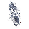 6jzkC C: citing same article ( M: map data used to model this data |
|---|---|
| Similar structure data | |
| EM raw data | EMPIAR-11015 (Title: Raw data for "Structure of polymerized type V pilin reveals assembly mechanism involving protease-mediated strand exchange" Data size: 72.1 Data #1: FimA motion-corrected dose-weighted frame sums [micrographs - single frame]) |


-Links
 PDBj
PDBj


- Assembly
| Deposited unit | 
|
|---|---|
| 1 |
|
-Components
| #1: Protein | Mass: 36488.805 Da / Num. of mol.: 4 Source method: isolated from a genetically manipulated source Source: (gene. exp.) Porphyromonas gingivalis ATCC 33277 (bacteria)Gene: fimA, PGN_0180 / Production host: Escherichia coli BL21 (bacteria) / References: UniProt: B2RH54 |
|---|
-Experimental details
-Experiment
| Experiment | Method: ELECTRON MICROSCOPY |
|---|---|
| EM experiment | Aggregation state: FILAMENT / 3D reconstruction method: single particle reconstruction |
- Sample preparation
| Component | Name: Type V pilus (FimA) / Type: COMPLEX / Entity ID: all / Source: RECOMBINANT |
|---|---|
| Molecular weight | Value: 6 kDa/nm / Experimental value: NO |
| Source (natural) | Organism: Porphyromonas gingivalis ATCC 33277 (bacteria) |
| Source (recombinant) | Organism: Escherichia coli BL21 (bacteria) / Strain: BL21 |
| Buffer solution | pH: 7.5 |
| Buffer component | Conc.: 20 mM / Name: Tris / Formula: C4H11NO3 |
| Specimen | Conc.: 1.5 mg/ml / Embedding applied: NO / Shadowing applied: NO / Staining applied: NO / Vitrification applied: YES / Details: matured, polymerized state |
| Specimen support | Details: Gatan Solarus / Grid material: COPPER / Grid mesh size: 400 divisions/in. / Grid type: Quantifoil R1.2/1.3 |
| Vitrification | Instrument: FEI VITROBOT MARK IV / Cryogen name: ETHANE-PROPANE / Humidity: 100 % / Chamber temperature: 289 K / Details: 3 second blot, 3.0uL |
- Electron microscopy imaging
| Experimental equipment |  Model: Talos Arctica / Image courtesy: FEI Company |
|---|---|
| Microscopy | Model: FEI TALOS ARCTICA / Details: nanoprobe, parallel beam illumination |
| Electron gun | Electron source: FIELD EMISSION GUN / Accelerating voltage: 200 kV / Illumination mode: FLOOD BEAM |
| Electron lens | Mode: BRIGHT FIELDBright-field microscopy / Nominal magnification: 92000 X / Calibrated magnification: 125000 X / Nominal defocus max: -2500 nm / Nominal defocus min: -1500 nm / Cs: 2.7 mm / C2 aperture diameter: 30 µm / Alignment procedure: COMA FREE |
| Specimen holder | Cryogen: NITROGEN / Specimen holder model: FEI TITAN KRIOS AUTOGRID HOLDER / Temperature (max): 100 K / Temperature (min): 77 K / Residual tilt: 0.1 mradians |
| Image recording | Average exposure time: 60 sec. / Electron dose: 46 e/Å2 / Detector mode: COUNTING / Film or detector model: FEI FALCON III (4k x 4k) / Num. of grids imaged: 1 / Num. of real images: 1153 Details: frame alignment and dose weighting using motioncor2 |
| Image scans | Sampling size: 14 µm / Width: 4000 / Height: 4000 / Movie frames/image: 75 / Used frames/image: 1-75 |
- Processing
| EM software |
| |||||||||||||||||||||||||||||||||||||||||||||
|---|---|---|---|---|---|---|---|---|---|---|---|---|---|---|---|---|---|---|---|---|---|---|---|---|---|---|---|---|---|---|---|---|---|---|---|---|---|---|---|---|---|---|---|---|---|---|
| Image processing | Details: frame alignment and integration with motioncor2 incl. dose weighting | |||||||||||||||||||||||||||||||||||||||||||||
| CTF correction | Type: PHASE FLIPPING AND AMPLITUDE CORRECTION | |||||||||||||||||||||||||||||||||||||||||||||
| Particle selection | Num. of particles selected: 831459 | |||||||||||||||||||||||||||||||||||||||||||||
| Symmetry | Point symmetry: C1 (asymmetric) | |||||||||||||||||||||||||||||||||||||||||||||
| 3D reconstruction | Resolution: 3.6 Å / Resolution method: FSC 0.143 CUT-OFF / Num. of particles: 61728 / Algorithm: FOURIER SPACE / Details: independent / Num. of class averages: 1 / Symmetry type: POINT | |||||||||||||||||||||||||||||||||||||||||||||
| Atomic model building | B value: 128 / Protocol: FLEXIBLE FIT / Space: REAL / Target criteria: CC | |||||||||||||||||||||||||||||||||||||||||||||
| Atomic model building | PDB-ID: 6JZJ Pdb chain-ID: A / Accession code: 6JZJ / Source name: PDB / Type: experimental model |
