 Movie
Movie Controller

[English] 日本語
 Yorodumi
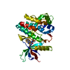
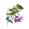
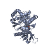
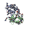
- PDB-6k93: Crystal structure of the type III effector XopAI from Xanthomonas... -
+ Open data

- Basic information
| Entry | Database: PDB / ID: 6k93 | ||||||
|---|---|---|---|---|---|---|---|
| Title | Crystal structure of the type III effector XopAI from Xanthomonas axonopodis pv. citri in space group P41212 | ||||||
 Components Components | Type III effector XopAI | ||||||
 Keywords Keywords | CELL INVASION / type III effectors / peptide-binding domain / mono-ADP-ribosyltransferase fold /  Xanthomonas axonopodis pv. citri Xanthomonas axonopodis pv. citri | ||||||
| Function / homology |  Function and homology information Function and homology informationcellular anatomical entity / NAD+-protein-arginine ADP-ribosyltransferase / NAD+-protein-arginine ADP-ribosyltransferase activity / nucleotidyltransferase activitySimilarity search - Function | ||||||
| Biological species |  Xanthomonas citri (bacteria) Xanthomonas citri (bacteria) | ||||||
| Method | X-RAY DIFFRACTION / SYNCHROTRON / MOLECULAR REPLACEMENT / Resolution: 1.53 Å | ||||||
 Authors Authors | Liu, J.-H. / Lai, Y.H. / Yang, J.-Y. / Hou, M.-H. | ||||||
| Funding support |  Taiwan, 1items Taiwan, 1items
| ||||||
 Citation Citation | Journal: Int J Mol Sci / Year: 2019 Title: Crystal Structure-Based Exploration of Arginine-Containing Peptide Binding in the ADP-Ribosyltransferase Domain of the Type III Effector XopAI Protein. Authors: Liu, J.H. / Yang, J.Y. / Hsu, D.W. / Lai, Y.H. / Li, Y.P. / Tsai, Y.R. / Hou, M.H. | ||||||
| History |
|
- Structure visualization
| Structure viewer | Molecule: MolmilJmol/JSmol |
|---|
- Downloads & links
-Download
| PDBx/mmCIF format | 6k93.cif.gz | 168.9 KB | Display | PDBx/mmCIF format |
|---|---|---|---|---|
| PDB format | pdb6k93.ent.gz | 129.5 KB | Display | PDB format |
| PDBx/mmJSON format | 6k93.json.gz | Tree view | PDBx/mmJSON format | |
| Others |  Other downloads Other downloads |
-Validation report
| Arichive directory | https://data.pdbj.org/pub/pdb/validation_reports/k9/6k93ftp://data.pdbj.org/pub/pdb/validation_reports/k9/6k93 | HTTPS FTP |
|---|
-Related structure data
| Related structure data | 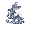 6k94C 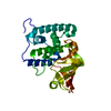 6klyC  4eln C: citing same article ( S: Starting model for refinement |
|---|---|
| Similar structure data |


-Links
 PDBj
PDBj- Assembly
| Deposited unit | 
| |||||||||||||||
|---|---|---|---|---|---|---|---|---|---|---|---|---|---|---|---|---|
| 1 |
| |||||||||||||||
| Unit cell |
| |||||||||||||||
| Components on special symmetry positions |
|
-Components
| #1: Protein | Mass: 35866.039 Da / Num. of mol.: 1 Source method: isolated from a genetically manipulated source Source: (gene. exp.) Xanthomonas citri (bacteria) / Strain: XW19 / Gene: XopAI, XAC3230 / Plasmid: pET28a / Production host: Escherichia coli BL21(DE3) (bacteria) / Strain (production host): BL21(DE3) / Variant (production host): RIL+ / References: UniProt: Q8PHM1 |
|---|---|
| #2: Water | ChemComp-HOH / Water Mass: 18.015 Da / Num. of mol.: 374 / Source method: isolated from a natural source / Formula: H2O Mass: 18.015 Da / Num. of mol.: 374 / Source method: isolated from a natural source / Formula: H2O |
-Experimental details
-Experiment
| Experiment | Method: X-RAY DIFFRACTION / Number of used crystals: 1 |
|---|
- Sample preparation
| Crystal | Density Matthews: 2.07 Å3/Da / Density % sol: 40.71 % |
|---|---|
| Crystal grow | Temperature: 298 K / Method: vapor diffusion, hanging drop / pH: 8 Details: 16.5% 7(V/V) PEG400, 6.5%(W/V) PEG 20000, 50mM KH2PO4, pH 8.0, 1 uL of 10 mM beta-mercaptoethanol |
-Data collection
| Diffraction | Mean temperature: 110 K / Serial crystal experiment: N |
|---|---|
| Diffraction source | Source: SYNCHROTRON / Site: NSRRC / Beamline: TPS 05A / Wavelength: 0.9198 Å |
| Detector | Type: RAYONIX MX300-HS / Detector: CCD / Date: Nov 4, 2016 |
| Radiation | Protocol: SINGLE WAVELENGTH / Monochromatic (M) / Laue (L): M / Scattering type: x-ray |
| Radiation wavelength | Wavelength: 0.9198 Å / Relative weight: 1 |
| Reflection | Resolution: 1.53→37.48 Å / Num. obs: 47011 / % possible obs: 100 % / Redundancy: 10.9 % / Biso Wilson estimate: 14.597 Å2 / CC1/2: 0.997 / Rmerge(I) obs: 0.131 / Rpim(I) all: 0.041 / Rrim(I) all: 0.138 / Χ2: 0.91 / Net I/σ(I): 9.6 |
| Reflection shell | Resolution: 1.53→1.56 Å / Redundancy: 6.4 % / Rmerge(I) obs: 1.064 / Num. unique obs: 2290 / CC1/2: 0.647 / Rpim(I) all: 0.464 / Χ2: 0.66 / % possible all: 100 |
- Processing
| Software |
| ||||||||||||||||||||||||||||||||||||||||||||||||||||||||||||||||||||||||||||||||||||||||||||||||||||||||||||||||||||||||||||||||||||||||||||||||||||||||||||||||||||||||||||||||||||||||||||||||||||||||||||||||||||||||||||||||||||||||||||||||||||||||||
|---|---|---|---|---|---|---|---|---|---|---|---|---|---|---|---|---|---|---|---|---|---|---|---|---|---|---|---|---|---|---|---|---|---|---|---|---|---|---|---|---|---|---|---|---|---|---|---|---|---|---|---|---|---|---|---|---|---|---|---|---|---|---|---|---|---|---|---|---|---|---|---|---|---|---|---|---|---|---|---|---|---|---|---|---|---|---|---|---|---|---|---|---|---|---|---|---|---|---|---|---|---|---|---|---|---|---|---|---|---|---|---|---|---|---|---|---|---|---|---|---|---|---|---|---|---|---|---|---|---|---|---|---|---|---|---|---|---|---|---|---|---|---|---|---|---|---|---|---|---|---|---|---|---|---|---|---|---|---|---|---|---|---|---|---|---|---|---|---|---|---|---|---|---|---|---|---|---|---|---|---|---|---|---|---|---|---|---|---|---|---|---|---|---|---|---|---|---|---|---|---|---|---|---|---|---|---|---|---|---|---|---|---|---|---|---|---|---|---|---|---|---|---|---|---|---|---|---|---|---|---|---|---|---|---|---|---|---|---|---|---|---|---|---|---|---|---|---|---|---|---|---|
| Refinement | Method to determine structure: MOLECULAR REPLACEMENT Starting model: 4eln 4eln Resolution: 1.53→36.891 Å / Cross valid method: FREE R-VALUE / Phase error: 16.04
| ||||||||||||||||||||||||||||||||||||||||||||||||||||||||||||||||||||||||||||||||||||||||||||||||||||||||||||||||||||||||||||||||||||||||||||||||||||||||||||||||||||||||||||||||||||||||||||||||||||||||||||||||||||||||||||||||||||||||||||||||||||||||||
| Solvent computation | Shrinkage radii: 0.9 Å / VDW probe radii: 1.11 Å | ||||||||||||||||||||||||||||||||||||||||||||||||||||||||||||||||||||||||||||||||||||||||||||||||||||||||||||||||||||||||||||||||||||||||||||||||||||||||||||||||||||||||||||||||||||||||||||||||||||||||||||||||||||||||||||||||||||||||||||||||||||||||||
| Displacement parameters | Biso mean: 22.65 Å2 | ||||||||||||||||||||||||||||||||||||||||||||||||||||||||||||||||||||||||||||||||||||||||||||||||||||||||||||||||||||||||||||||||||||||||||||||||||||||||||||||||||||||||||||||||||||||||||||||||||||||||||||||||||||||||||||||||||||||||||||||||||||||||||
| Refinement step | Cycle: LAST / Resolution: 1.53→36.891 Å
| ||||||||||||||||||||||||||||||||||||||||||||||||||||||||||||||||||||||||||||||||||||||||||||||||||||||||||||||||||||||||||||||||||||||||||||||||||||||||||||||||||||||||||||||||||||||||||||||||||||||||||||||||||||||||||||||||||||||||||||||||||||||||||
| Refinement TLS params. | Method: refined / Refine-ID: X-RAY DIFFRACTION
| ||||||||||||||||||||||||||||||||||||||||||||||||||||||||||||||||||||||||||||||||||||||||||||||||||||||||||||||||||||||||||||||||||||||||||||||||||||||||||||||||||||||||||||||||||||||||||||||||||||||||||||||||||||||||||||||||||||||||||||||||||||||||||
| Refinement TLS group |
|
