 Movie
Movie Controller

[English] 日本語
 Yorodumi
- PDB-6jwg: Crystal structure of Formate dehydrogenase mutant C256I/E261P/S38... -
+ Open data

- Basic information
| Entry | Database: PDB / ID: 6jwg | ||||||
|---|---|---|---|---|---|---|---|
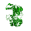
| Title | Crystal structure of Formate dehydrogenase mutant C256I/E261P/S381I from Pseudomonas sp. 101 | ||||||
 Components Components | Formate dehydrogenase | ||||||
 Keywords Keywords | OXIDOREDUCTASE / Formate dehydrogenase | ||||||
| Function / homology |  Function and homology information Function and homology informationformate catabolic process / formate dehydrogenase / formate dehydrogenase (NAD+) activity / oxidoreductase activity, acting on the CH-OH group of donors, NAD or NADP as acceptor / NAD binding / cytoplasmSimilarity search - Function | ||||||
| Biological species |  Pseudomonas sp. 101 (bacteria) Pseudomonas sp. 101 (bacteria) | ||||||
| Method | X-RAY DIFFRACTION / SYNCHROTRON / MOLECULAR REPLACEMENT / Resolution: 2.081 Å | ||||||
 Authors Authors | Feng, Y. / Guo, X. / Xue, S. / Zhao, Z. | ||||||
 Citation Citation | Journal: Chemistry / Year: 2020 Title: Structure-Guided Design of Formate Dehydrogenase for Regeneration of a Non-Natural Redox Cofactor. Authors: Guo, X. / Wang, X. / Liu, Y. / Li, Q. / Wang, J. / Liu, W. / Zhao, Z.K. | ||||||
| History |
|
- Structure visualization
| Structure viewer | Molecule: MolmilJmol/JSmol |
|---|
- Downloads & links
-Download
| PDBx/mmCIF format | 6jwg.cif.gz | 173.5 KB | Display | PDBx/mmCIF format |
|---|---|---|---|---|
| PDB format | pdb6jwg.ent.gz | 135.1 KB | Display | PDB format |
| PDBx/mmJSON format | 6jwg.json.gz | Tree view | PDBx/mmJSON format | |
| Others |  Other downloads Other downloads |
-Validation report
| Arichive directory | https://data.pdbj.org/pub/pdb/validation_reports/jw/6jwgftp://data.pdbj.org/pub/pdb/validation_reports/jw/6jwg | HTTPS FTP |
|---|
-Related structure data
| Related structure data | 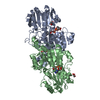 6jujC 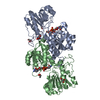 6jukC 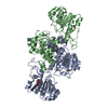 6jx1C 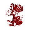 2go1S S: Starting model for refinement C: citing same article ( |
|---|---|
| Similar structure data |


-Links
 PDBj
PDBj- Assembly
| Deposited unit | 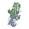
| ||||||||
|---|---|---|---|---|---|---|---|---|---|
| 1 |
| ||||||||
| Unit cell |
| ||||||||
| Components on special symmetry positions |
|
-Components
| #1: Protein | / FDH / NAD-dependent formate dehydrogenase Mass: 44198.184 Da / Num. of mol.: 2 / Mutation: C256I/E261P/S381I Source method: isolated from a genetically manipulated source Source: (gene. exp.) Pseudomonas sp. 101 (bacteria) / Production host: Escherichia coli BL21(DE3) (bacteria) / Strain (production host): BL21(DE3) / References: UniProt: P33160, formate dehydrogenase#2: Chemical | Glycerol  Mass: 92.094 Da / Num. of mol.: 2 / Source method: obtained synthetically / Formula: C3H8O3 Mass: 92.094 Da / Num. of mol.: 2 / Source method: obtained synthetically / Formula: C3H8O3#3: Chemical | ChemComp-TRS / | Tris  Mass: 122.143 Da / Num. of mol.: 1 / Source method: obtained synthetically / Formula: C4H12NO3 / Comment: pH buffer*YM Mass: 122.143 Da / Num. of mol.: 1 / Source method: obtained synthetically / Formula: C4H12NO3 / Comment: pH buffer*YM#4: Water | ChemComp-HOH / | Water Mass: 18.015 Da / Num. of mol.: 685 / Source method: isolated from a natural source / Formula: H2O Mass: 18.015 Da / Num. of mol.: 685 / Source method: isolated from a natural source / Formula: H2O |
|---|
-Experimental details
-Experiment
| Experiment | Method: X-RAY DIFFRACTION / Number of used crystals: 1 |
|---|
- Sample preparation
| Crystal | Density Matthews: 2.65 Å3/Da / Density % sol: 53.67 % |
|---|---|
| Crystal grow | Temperature: 300 K / Method: vapor diffusion, hanging drop / pH: 6.5 Details: 0.2 M Ammonium sulfate, 0.1 M MES monohydrate pH 6.5, 30% w/v Polyethylene glycol monomethyl ether 5,000 |
-Data collection
| Diffraction | Mean temperature: 100 K / Serial crystal experiment: N |
|---|---|
| Diffraction source | Source: SYNCHROTRON / Site: SSRF  / Beamline: BL19U1 / Wavelength: 0.9793 Å / Beamline: BL19U1 / Wavelength: 0.9793 Å |
| Detector | Type: MAR CCD 130 mm / Detector: CCD / Date: Dec 28, 2017 |
| Radiation | Protocol: SINGLE WAVELENGTH / Monochromatic (M) / Laue (L): M / Scattering type: x-ray |
| Radiation wavelength | Wavelength: 0.9793 Å / Relative weight: 1 |
| Reflection | Resolution: 2.08→50 Å / Num. obs: 53181 / % possible obs: 95.98 % / Redundancy: 5.6 % / Biso Wilson estimate: 25.03 Å2 / CC1/2: 0.974 / Rmerge(I) obs: 0.11 / Net I/σ(I): 13.86 |
| Reflection shell | Resolution: 2.08→2.16 Å / Redundancy: 5.1 % / Rmerge(I) obs: 0.51 / Mean I/σ(I) obs: 2.86 / Num. unique obs: 3759 / CC1/2: 0.881 / % possible all: 68.53 |
- Processing
| Software |
| |||||||||||||||||||||||||||||||||||||||||||||||||||||||||||||||||||||||||||||||||||||||||||||||||||||||||
|---|---|---|---|---|---|---|---|---|---|---|---|---|---|---|---|---|---|---|---|---|---|---|---|---|---|---|---|---|---|---|---|---|---|---|---|---|---|---|---|---|---|---|---|---|---|---|---|---|---|---|---|---|---|---|---|---|---|---|---|---|---|---|---|---|---|---|---|---|---|---|---|---|---|---|---|---|---|---|---|---|---|---|---|---|---|---|---|---|---|---|---|---|---|---|---|---|---|---|---|---|---|---|---|---|---|---|
| Refinement | Method to determine structure: MOLECULAR REPLACEMENT Starting model: 2GO1 Resolution: 2.081→44.062 Å / SU ML: 0.16 / Cross valid method: FREE R-VALUE / σ(F): 1.35 / Phase error: 18.96
| |||||||||||||||||||||||||||||||||||||||||||||||||||||||||||||||||||||||||||||||||||||||||||||||||||||||||
| Solvent computation | Shrinkage radii: 0.9 Å / VDW probe radii: 1.11 Å | |||||||||||||||||||||||||||||||||||||||||||||||||||||||||||||||||||||||||||||||||||||||||||||||||||||||||
| Refinement step | Cycle: LAST / Resolution: 2.081→44.062 Å
| |||||||||||||||||||||||||||||||||||||||||||||||||||||||||||||||||||||||||||||||||||||||||||||||||||||||||
| Refine LS restraints |
| |||||||||||||||||||||||||||||||||||||||||||||||||||||||||||||||||||||||||||||||||||||||||||||||||||||||||
| LS refinement shell |
|
