 Movie
Movie Controller

[English] 日本語
 Yorodumi
- PDB-6ju1: p-Hydroxybenzoate hydroxylase Y385F mutant complexed with 3,4-dih... -
+ Open data

- Basic information
| Entry | Database: PDB / ID: 6ju1 | ||||||
|---|---|---|---|---|---|---|---|
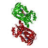
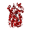
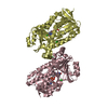
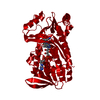
| Title | p-Hydroxybenzoate hydroxylase Y385F mutant complexed with 3,4-dihydroxybenzoate | ||||||
 Components Components | 4-hydroxybenzoate 3-monooxygenase | ||||||
 Keywords Keywords | OXIDOREDUCTASE / Aromatic compound hydroxylase / flavin / FAD | ||||||
| Function / homology |  Function and homology information Function and homology informationbenzoate catabolic process / 4-hydroxybenzoate 3-monooxygenase (NADPH) activity / 4-hydroxybenzoate 3-monooxygenase / 4-hydroxybenzoate 3-monooxygenase activity / benzoate catabolic process via hydroxylation / FAD binding / flavin adenine dinucleotide binding / oxidoreductase activitySimilarity search - Function | ||||||
| Biological species |   Pseudomonas aeruginosa (bacteria) Pseudomonas aeruginosa (bacteria) | ||||||
| Method | X-RAY DIFFRACTION / SYNCHROTRON / FOURIER SYNTHESIS / Resolution: 1.6 Å | ||||||
 Authors Authors | Yato, M. / Arakawa, T. / Yamada, C. / Fushinobu, S. | ||||||
 Citation Citation | Journal: Biochemistry / Year: 2019 Title: Understanding the Molecular Mechanism Underlying the High Catalytic Activity ofp-Hydroxybenzoate Hydroxylase Mutants for Producing Gallic Acid. Authors: Moriwaki, Y. / Yato, M. / Terada, T. / Saito, S. / Nukui, N. / Iwasaki, T. / Nishi, T. / Kawaguchi, Y. / Okamoto, K. / Arakawa, T. / Yamada, C. / Fushinobu, S. / Shimizu, K. | ||||||
| History |
|
- Structure visualization
| Structure viewer | Molecule: MolmilJmol/JSmol |
|---|
- Downloads & links
-Download
| PDBx/mmCIF format | 6ju1.cif.gz | 110.8 KB | Display | PDBx/mmCIF format |
|---|---|---|---|---|
| PDB format | pdb6ju1.ent.gz | 79.5 KB | Display | PDB format |
| PDBx/mmJSON format | 6ju1.json.gz | Tree view | PDBx/mmJSON format | |
| Others |  Other downloads Other downloads |
-Validation report
| Arichive directory | https://data.pdbj.org/pub/pdb/validation_reports/ju/6ju1ftp://data.pdbj.org/pub/pdb/validation_reports/ju/6ju1 | HTTPS FTP |
|---|
-Related structure data
| Related structure data | 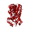 1iuvS S: Starting model for refinement |
|---|---|
| Similar structure data |
-Links
 PDBj
PDBj

- Assembly
| Deposited unit | 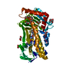
| ||||||||||||
|---|---|---|---|---|---|---|---|---|---|---|---|---|---|
| 1 | 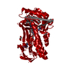
| ||||||||||||
| Unit cell |
| ||||||||||||
| Components on special symmetry positions |
|
-Components
-Protein , 1 types, 1 molecules A
| #1: Protein | Mass: 46537.914 Da / Num. of mol.: 1 / Mutation: Y385F Source method: isolated from a genetically manipulated source Source: (gene. exp.) Pseudomonas aeruginosa (bacteria) / Gene: pobA, DT376_03235 / Plasmid: pET-28b / Production host: Escherichia coli (E. coli) / Strain (production host): BL21-CodonPlus (DE3)-RILReferences: UniProt: A0A367MFY2, UniProt: P20586*PLUS, 4-hydroxybenzoate 3-monooxygenase |
|---|
-Non-polymers , 7 types, 408 molecules 


| #2: Chemical | ChemComp-FAD / Flavin adenine dinucleotide Mass: 785.550 Da / Num. of mol.: 1 / Source method: obtained synthetically / Formula: C27H33N9O15P2 / Feature type: SUBJECT OF INVESTIGATION / Comment: FAD*YM Mass: 785.550 Da / Num. of mol.: 1 / Source method: obtained synthetically / Formula: C27H33N9O15P2 / Feature type: SUBJECT OF INVESTIGATION / Comment: FAD*YM | ||||||||
|---|---|---|---|---|---|---|---|---|---|
| #3: Chemical | ChemComp-DHB / Protocatechuic acid Mass: 154.120 Da / Num. of mol.: 1 / Source method: obtained synthetically / Formula: C7H6O4 / Feature type: SUBJECT OF INVESTIGATION Mass: 154.120 Da / Num. of mol.: 1 / Source method: obtained synthetically / Formula: C7H6O4 / Feature type: SUBJECT OF INVESTIGATION | ||||||||
| #4: Chemical | ChemComp-GOL / Glycerol Mass: 92.094 Da / Num. of mol.: 4 / Source method: obtained synthetically / Formula: C3H8O3 Mass: 92.094 Da / Num. of mol.: 4 / Source method: obtained synthetically / Formula: C3H8O3#5: Chemical | ChemComp-PG4 / | Polyethylene glycol Mass: 194.226 Da / Num. of mol.: 1 / Source method: obtained synthetically / Formula: C8H18O5 / Comment: precipitant*YM Mass: 194.226 Da / Num. of mol.: 1 / Source method: obtained synthetically / Formula: C8H18O5 / Comment: precipitant*YM#6: Chemical | ChemComp-P33 / | Polyethylene glycol Mass: 326.383 Da / Num. of mol.: 1 / Source method: obtained synthetically / Formula: C14H30O8 / Comment: precipitant*YM Mass: 326.383 Da / Num. of mol.: 1 / Source method: obtained synthetically / Formula: C14H30O8 / Comment: precipitant*YM#7: Chemical | Bromide Mass: 79.904 Da / Num. of mol.: 2 / Source method: obtained synthetically / Formula: Br Mass: 79.904 Da / Num. of mol.: 2 / Source method: obtained synthetically / Formula: Br#8: Water | ChemComp-HOH / | WaterMass: 18.015 Da / Num. of mol.: 398 / Source method: isolated from a natural source / Formula: H2O |
-Experimental details
-Experiment
| Experiment | Method: X-RAY DIFFRACTION / Number of used crystals: 1 |
|---|
- Sample preparation
| Crystal | Density Matthews: 2.4 Å3/Da / Density % sol: 48.75 % |
|---|---|
| Crystal grow | Temperature: 293.15 K / Method: vapor diffusion, sitting drop / pH: 7.5 / Details: 0.15 M KBr, 30% PEG 2000 monomethyl ether |
-Data collection
| Diffraction | Mean temperature: 100 K / Serial crystal experiment: N |
|---|---|
| Diffraction source | Source: SYNCHROTRON / Site: Photon Factory  / Beamline: AR-NW12A / Wavelength: 1.29 Å / Beamline: AR-NW12A / Wavelength: 1.29 Å |
| Detector | Type: DECTRIS PILATUS3 S 2M / Detector: PIXEL / Date: Dec 14, 2018 |
| Radiation | Monochromator: Numerical link type Si(111) double crystal / Protocol: SINGLE WAVELENGTH / Monochromatic (M) / Laue (L): M / Scattering type: x-ray |
| Radiation wavelength | Wavelength: 1.29 Å / Relative weight: 1 |
| Reflection | Resolution: 1.6→50 Å / Num. obs: 59096 / % possible obs: 99.6 % / Redundancy: 12.4 % / Rmerge(I) obs: 0.103 / Net I/σ(I): 28.1 |
| Reflection shell | Resolution: 1.6→1.63 Å / Redundancy: 7.8 % / Rmerge(I) obs: 0.914 / Mean I/σ(I) obs: 1.3 / CC1/2: 0.751 / % possible all: 93 |
- Processing
| Software |
| ||||||||||||||||||||||||||||||||||||||||||||||||||||||||||||||||||||||||||||||||||||||||||||||||||||||||||||||||||||||||||||||||||||||||||||||||||||||||||||||||||||||||||||||||||||||
|---|---|---|---|---|---|---|---|---|---|---|---|---|---|---|---|---|---|---|---|---|---|---|---|---|---|---|---|---|---|---|---|---|---|---|---|---|---|---|---|---|---|---|---|---|---|---|---|---|---|---|---|---|---|---|---|---|---|---|---|---|---|---|---|---|---|---|---|---|---|---|---|---|---|---|---|---|---|---|---|---|---|---|---|---|---|---|---|---|---|---|---|---|---|---|---|---|---|---|---|---|---|---|---|---|---|---|---|---|---|---|---|---|---|---|---|---|---|---|---|---|---|---|---|---|---|---|---|---|---|---|---|---|---|---|---|---|---|---|---|---|---|---|---|---|---|---|---|---|---|---|---|---|---|---|---|---|---|---|---|---|---|---|---|---|---|---|---|---|---|---|---|---|---|---|---|---|---|---|---|---|---|---|---|
| Refinement | Method to determine structure: FOURIER SYNTHESIS Starting model: 1IUV Resolution: 1.6→40.13 Å / Cor.coef. Fo:Fc: 0.974 / Cor.coef. Fo:Fc free: 0.967 / SU B: 1.814 / SU ML: 0.06 / Cross valid method: THROUGHOUT / ESU R: 0.076 / ESU R Free: 0.077 / Details: HYDROGENS HAVE BEEN ADDED IN THE RIDING POSITIONS
| ||||||||||||||||||||||||||||||||||||||||||||||||||||||||||||||||||||||||||||||||||||||||||||||||||||||||||||||||||||||||||||||||||||||||||||||||||||||||||||||||||||||||||||||||||||||
| Solvent computation | Ion probe radii: 0.8 Å / Shrinkage radii: 0.8 Å / VDW probe radii: 1.2 Å | ||||||||||||||||||||||||||||||||||||||||||||||||||||||||||||||||||||||||||||||||||||||||||||||||||||||||||||||||||||||||||||||||||||||||||||||||||||||||||||||||||||||||||||||||||||||
| Displacement parameters | Biso mean: 23.047 Å2
| ||||||||||||||||||||||||||||||||||||||||||||||||||||||||||||||||||||||||||||||||||||||||||||||||||||||||||||||||||||||||||||||||||||||||||||||||||||||||||||||||||||||||||||||||||||||
| Refinement step | Cycle: 1 / Resolution: 1.6→40.13 Å
| ||||||||||||||||||||||||||||||||||||||||||||||||||||||||||||||||||||||||||||||||||||||||||||||||||||||||||||||||||||||||||||||||||||||||||||||||||||||||||||||||||||||||||||||||||||||
| Refine LS restraints |
|