 Movie
Movie Controller

[English] 日本語
 Yorodumi
- PDB-6jls: Crystal Structure of FMN-dependent Cysteine Decarboxylases TvaF f... -
+ Open data

- Basic information
| Entry | Database: PDB / ID: 6jls | ||||||
|---|---|---|---|---|---|---|---|
| Title | Crystal Structure of FMN-dependent Cysteine Decarboxylases TvaF from Thioviridamide Biosynthesis | ||||||
 Components Components | Putative flavoprotein decarboxylase | ||||||
 Keywords Keywords |  BIOSYNTHETIC PROTEIN / FMN-dependent BIOSYNTHETIC PROTEIN / FMN-dependent | ||||||
| Function / homology | Flavoprotein / Flavin prenyltransferase-like / Flavoprotein / catalytic activity / FLAVIN MONONUCLEOTIDE / Phosphopantothenoylcysteine decarboxylase Function and homology information Function and homology information | ||||||
| Biological species |  Streptomyces olivoviridis (bacteria) Streptomyces olivoviridis (bacteria) | ||||||
| Method | X-RAY DIFFRACTION / SYNCHROTRON / MOLECULAR REPLACEMENT / Resolution: 2.24 Å | ||||||
 Authors Authors | Li, J. / Lu, J. / Wang, H. / Zhu, J. | ||||||
| Funding support |  China, 1items China, 1items
| ||||||
 Citation Citation | Journal: Org.Lett. / Year: 2019 Title: Characterization of the FMN-Dependent Cysteine Decarboxylase from Thioviridamide Biosynthesis. Authors: Lu, J. / Li, J. / Wu, Y. / Fang, X. / Zhu, J. / Wang, H. | ||||||
| History |
|
- Structure visualization
| Structure viewer | Molecule: MolmilJmol/JSmol |
|---|
- Downloads & links
-Download
| PDBx/mmCIF format | 6jls.cif.gz | 80.2 KB | Display | PDBx/mmCIF format |
|---|---|---|---|---|
| PDB format | pdb6jls.ent.gz | 59.9 KB | Display | PDB format |
| PDBx/mmJSON format | 6jls.json.gz | Tree view | PDBx/mmJSON format | |
| Others |  Other downloads Other downloads |
-Validation report
| Arichive directory | https://data.pdbj.org/pub/pdb/validation_reports/jl/6jlsftp://data.pdbj.org/pub/pdb/validation_reports/jl/6jls | HTTPS FTP |
|---|
-Related structure data
| Related structure data | 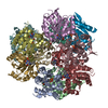 1g63S S: Starting model for refinement |
|---|---|
| Similar structure data |


-Links
 PDBj
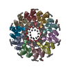
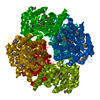
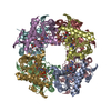
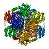
PDBj- Assembly
| Deposited unit | 
| ||||||||
|---|---|---|---|---|---|---|---|---|---|
| 1 | x 12
| ||||||||
| Unit cell |
| ||||||||
| Components on special symmetry positions |
|
-Components
| #1: Protein | Mass: 20404.195 Da / Num. of mol.: 1 Source method: isolated from a genetically manipulated source Source: (gene. exp.) Streptomyces olivoviridis (bacteria) / Gene: tvaF / Production host: Escherichia coli BL21 (bacteria) / Strain (production host): BL21 / References: UniProt: T2HUM4 |
|---|---|
| #2: Chemical | ChemComp-FMN / Flavin mononucleotide  Mass: 456.344 Da / Num. of mol.: 1 / Source method: obtained synthetically / Formula: C17H21N4O9P Mass: 456.344 Da / Num. of mol.: 1 / Source method: obtained synthetically / Formula: C17H21N4O9P |
| #3: Water | ChemComp-HOH / Water Mass: 18.015 Da / Num. of mol.: 62 / Source method: isolated from a natural source / Formula: H2O Mass: 18.015 Da / Num. of mol.: 62 / Source method: isolated from a natural source / Formula: H2O |
-Experimental details
-Experiment
| Experiment | Method: X-RAY DIFFRACTION / Number of used crystals: 1 |
|---|
- Sample preparation
| Crystal | Density Matthews: 5.23 Å3/Da / Density % sol: 76.51 % |
|---|---|
| Crystal grow | Temperature: 295 K / Method: vapor diffusion, sitting drop Details: Two micro liters of protein solution containing 7 mg/ml Tvaf in 20 mM Tris, pH 8.0, 50 mM NaCl, was mixed with 2 micro liters of well solution containing 0.1 M Sodium chloride, 0.1M Bicine ...Details: Two micro liters of protein solution containing 7 mg/ml Tvaf in 20 mM Tris, pH 8.0, 50 mM NaCl, was mixed with 2 micro liters of well solution containing 0.1 M Sodium chloride, 0.1M Bicine pH 9.0, and 30% PEG 500 MME (vol/vol). The crystallization drop was incubated against 100 micro liters of well solution. |
-Data collection
| Diffraction | Mean temperature: 100 K / Serial crystal experiment: N |
|---|---|
| Diffraction source | Source: SYNCHROTRON / Site: SSRF / Beamline: BL18U1 / Wavelength: 0.9793 Å |
| Detector | Type: DECTRIS PILATUS3 S 6M / Detector: PIXEL / Date: Oct 26, 2018 |
| Radiation | Protocol: SINGLE WAVELENGTH / Monochromatic (M) / Laue (L): M / Scattering type: x-ray |
| Radiation wavelength | Wavelength: 0.9793 Å / Relative weight: 1 |
| Reflection | Resolution: 2.24→76.691 Å / Num. obs: 21611 / % possible obs: 100 % / Redundancy: 29.8 % / CC1/2: 0.999 / Net I/σ(I): 11.5 |
| Reflection shell | Resolution: 2.24→2.31 Å / Num. unique obs: 1937 / CC1/2: 0.395 |
- Processing
| Software |
| |||||||||||||||||||||||||||||||||||||||||||||||||||||||||||||||
|---|---|---|---|---|---|---|---|---|---|---|---|---|---|---|---|---|---|---|---|---|---|---|---|---|---|---|---|---|---|---|---|---|---|---|---|---|---|---|---|---|---|---|---|---|---|---|---|---|---|---|---|---|---|---|---|---|---|---|---|---|---|---|---|---|
| Refinement | Method to determine structure: MOLECULAR REPLACEMENT Starting model: 1G63 Resolution: 2.24→76.691 Å / SU ML: 0.32 / Cross valid method: THROUGHOUT / σ(F): 1.39 / Phase error: 23.59 / Stereochemistry target values: ML
| |||||||||||||||||||||||||||||||||||||||||||||||||||||||||||||||
| Solvent computation | Shrinkage radii: 0.9 Å / VDW probe radii: 1.11 Å / Solvent model: FLAT BULK SOLVENT MODEL | |||||||||||||||||||||||||||||||||||||||||||||||||||||||||||||||
| Refinement step | Cycle: LAST / Resolution: 2.24→76.691 Å
| |||||||||||||||||||||||||||||||||||||||||||||||||||||||||||||||
| Refine LS restraints |
| |||||||||||||||||||||||||||||||||||||||||||||||||||||||||||||||
| LS refinement shell |
| |||||||||||||||||||||||||||||||||||||||||||||||||||||||||||||||
| Refinement TLS params. | Method: refined / Origin x: 83.5922 Å / Origin y: 69.3499 Å / Origin z: 49.3223 Å
| |||||||||||||||||||||||||||||||||||||||||||||||||||||||||||||||
| Refinement TLS group | Selection details: all |
