 Movie
Movie Controller

[English] 日本語
 Yorodumi
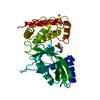
- PDB-6j7s: Crystal structure of toxin TglT (unusual type guanylyltransferase... -
+ Open data

- Basic information
| Entry | Database: PDB / ID: 6j7s | |||||||||
|---|---|---|---|---|---|---|---|---|---|---|
| Title | Crystal structure of toxin TglT (unusual type guanylyltransferase-like toxin, Rv1045) wild type protein from Mycobacterium tuberculosis | |||||||||
 Components Components | guanylyltransferase-like toxin | |||||||||
 Keywords Keywords |  TOXIN / guanylyltransferase / guanylyltransferase-like toxin / TA TOXIN / guanylyltransferase / guanylyltransferase-like toxin / TA | |||||||||
| Function / homology | Nucleotidyl transferase AbiEii toxin, Type IV TA system / Nucleotidyl transferase AbiEii toxin, Type IV TA system / Nucleotidyl transferase AbiEii/AbiGii toxin family protein Function and homology information Function and homology information | |||||||||
| Biological species |   Mycobacterium tuberculosis (bacteria) Mycobacterium tuberculosis (bacteria) | |||||||||
| Method | X-RAY DIFFRACTION / SYNCHROTRON / MOLECULAR REPLACEMENT / Resolution: 2.102 Å | |||||||||
 Authors Authors | Yu, X. / Gao, X. / Zhu, K. / Wojdyla, J.A. / Wang, M. / Cui, S. | |||||||||
| Funding support |  China, 2items China, 2items
| |||||||||
 Citation Citation | Journal: Commun Biol / Year: 2020 Title: Characterization of a toxin-antitoxin system in Mycobacterium tuberculosis suggests neutralization by phosphorylation as the antitoxicity mechanism. Authors: Yu, X. / Gao, X. / Zhu, K. / Yin, H. / Mao, X. / Wojdyla, J.A. / Qin, B. / Huang, H. / Wang, M. / Sun, Y.C. / Cui, S. | |||||||||
| History |
|
- Structure visualization
| Structure viewer | Molecule: MolmilJmol/JSmol |
|---|
- Downloads & links
-Download
| PDBx/mmCIF format | 6j7s.cif.gz | 123.6 KB | Display | PDBx/mmCIF format |
|---|---|---|---|---|
| PDB format | pdb6j7s.ent.gz | 95.2 KB | Display | PDB format |
| PDBx/mmJSON format | 6j7s.json.gz | Tree view | PDBx/mmJSON format | |
| Others |  Other downloads Other downloads |
-Validation report
| Arichive directory | https://data.pdbj.org/pub/pdb/validation_reports/j7/6j7sftp://data.pdbj.org/pub/pdb/validation_reports/j7/6j7s | HTTPS FTP |
|---|
-Related structure data
| Related structure data |  6j7nC  6j7oC  6j7pC  6j7qC  6j7rC  6j7tSC S: Starting model for refinement C: citing same article ( |
|---|---|
| Similar structure data |


-Links
 PDBj
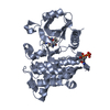
PDBj- Assembly
| Deposited unit | 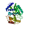
| ||||||||
|---|---|---|---|---|---|---|---|---|---|
| 1 |
| ||||||||
| Unit cell |
|
-Components
| #1: Protein | Mass: 34281.000 Da / Num. of mol.: 1 Source method: isolated from a genetically manipulated source Source: (gene. exp.) Mycobacterium tuberculosis (strain ATCC 25618 / H37Rv) (bacteria)Strain: ATCC 25618 / H37Rv / Gene: Rv1045 / Plasmid: pet28a / Production host: Escherichia coli BL21(DE3) (bacteria) / Strain (production host): BL21(DE3) / References: UniProt: P96356 | ||
|---|---|---|---|
| #2: Chemical | ChemComp-MG /   Mass: 24.305 Da / Num. of mol.: 13 / Source method: obtained synthetically / Formula: Mg Mass: 24.305 Da / Num. of mol.: 13 / Source method: obtained synthetically / Formula: Mg#3: Water | ChemComp-HOH / | Water Mass: 18.015 Da / Num. of mol.: 191 / Source method: isolated from a natural source / Formula: H2O Mass: 18.015 Da / Num. of mol.: 191 / Source method: isolated from a natural source / Formula: H2O |
-Experimental details
-Experiment
| Experiment | Method: X-RAY DIFFRACTION / Number of used crystals: 1 |
|---|
- Sample preparation
| Crystal | Density Matthews: 2.67 Å3/Da / Density % sol: 53.88 % |
|---|---|
| Crystal grow | Temperature: 296 K / Method: vapor diffusion, hanging drop / pH: 6.5 Details: 0.1 M Bis-Tris pH 6.5, 0.2 M magnesium chloride hexahydrate, 25% polyethylene glycol 3350 (v/v). |
-Data collection
| Diffraction | Mean temperature: 100 K / Serial crystal experiment: N |
|---|---|
| Diffraction source | Source: SYNCHROTRON / Site: SSRF / Beamline: BL19U1 / Wavelength: 0.9785 Å |
| Detector | Type: DECTRIS PILATUS3 S 6M / Detector: PIXEL / Date: Nov 19, 2017 |
| Radiation | Protocol: SINGLE WAVELENGTH / Monochromatic (M) / Laue (L): M / Scattering type: x-ray |
| Radiation wavelength | Wavelength: 0.9785 Å / Relative weight: 1 |
| Reflection | Resolution: 2.1→28.56 Å / Num. obs: 21592 / % possible obs: 99.9 % / Observed criterion σ(I): -3 / Redundancy: 11.04 % / Biso Wilson estimate: 39.74 Å2 / CC1/2: 0.999 / Rmerge(I) obs: 0.081 / Net I/σ(I): 23.47 |
| Reflection shell | Resolution: 2.1→2.23 Å / Redundancy: 11.23 % / Rmerge(I) obs: 0.746 / Mean I/σ(I) obs: 3.18 / Num. unique obs: 3430 / CC1/2: 0.905 / % possible all: 99.9 |
- Processing
| Software |
| |||||||||||||||||||||||||||||||||||||||||||||||||||||||||||||||
|---|---|---|---|---|---|---|---|---|---|---|---|---|---|---|---|---|---|---|---|---|---|---|---|---|---|---|---|---|---|---|---|---|---|---|---|---|---|---|---|---|---|---|---|---|---|---|---|---|---|---|---|---|---|---|---|---|---|---|---|---|---|---|---|---|
| Refinement | Method to determine structure: MOLECULAR REPLACEMENT Starting model: 6J7T Resolution: 2.102→28.56 Å / SU ML: 0.25 / Cross valid method: THROUGHOUT / σ(F): 1.34 / Phase error: 22.31
| |||||||||||||||||||||||||||||||||||||||||||||||||||||||||||||||
| Solvent computation | Shrinkage radii: 0.9 Å / VDW probe radii: 1.11 Å | |||||||||||||||||||||||||||||||||||||||||||||||||||||||||||||||
| Displacement parameters | Biso mean: 45.95 Å2 | |||||||||||||||||||||||||||||||||||||||||||||||||||||||||||||||
| Refinement step | Cycle: LAST / Resolution: 2.102→28.56 Å
| |||||||||||||||||||||||||||||||||||||||||||||||||||||||||||||||
| Refine LS restraints |
| |||||||||||||||||||||||||||||||||||||||||||||||||||||||||||||||
| LS refinement shell |
|
