 Movie
Movie Controller


+ Open data

- Basic information
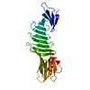
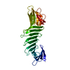
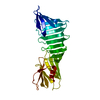
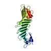
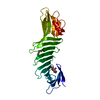
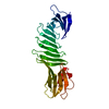
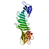
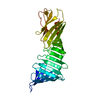
| Entry | Database: PDB / ID: 6j49 | |||||||||
|---|---|---|---|---|---|---|---|---|---|---|
| Title | Grafting VLADV sequence into OspAsm1 | |||||||||
 Components Components | Outer surface protein A | |||||||||
 Keywords Keywords | LIPID BINDING PROTEIN /  beta-sheet / DE NOVO PROTEIN beta-sheet / DE NOVO PROTEIN | |||||||||
| Function / homology |  Function and homology information Function and homology information | |||||||||
| Biological species |  Borrelia burgdorferi (Lyme disease spirochete) Borrelia burgdorferi (Lyme disease spirochete) | |||||||||
| Method | X-RAY DIFFRACTION / SYNCHROTRON / MOLECULAR REPLACEMENT / Resolution: 1.6 Å | |||||||||
 Authors Authors | Makabe, K. / Hori, Y. | |||||||||
 Citation Citation | Journal: Proteins / Year: 2019 Title: Grafting a short chameleon sequence from alpha B crystallin into a beta-sheet scaffold protein. Authors: Hori, Y. / Fujiwara, H. / Fujiwara, W. / Makabe, K. | |||||||||
| History |
|
- Structure visualization
| Structure viewer | Molecule: MolmilJmol/JSmol |
|---|
- Downloads & links
-Download
| PDBx/mmCIF format | 6j49.cif.gz | 68.8 KB | Display | PDBx/mmCIF format |
|---|---|---|---|---|
| PDB format | pdb6j49.ent.gz | 47.6 KB | Display | PDB format |
| PDBx/mmJSON format | 6j49.json.gz | Tree view | PDBx/mmJSON format | |
| Others |  Other downloads Other downloads |
-Validation report
| Arichive directory | https://data.pdbj.org/pub/pdb/validation_reports/j4/6j49ftp://data.pdbj.org/pub/pdb/validation_reports/j4/6j49 | HTTPS FTP |
|---|
-Related structure data
| Related structure data |  6j47C 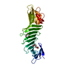 6j48C  2g8cS S: Starting model for refinement C: citing same article ( |
|---|---|
| Similar structure data |


-Links
 PDBj
PDBj


- Assembly
| Deposited unit | 
| ||||||||
|---|---|---|---|---|---|---|---|---|---|
| 1 |
| ||||||||
| Unit cell |
|
-Components
| #1: Protein | Mass: 26359.568 Da / Num. of mol.: 1 Mutation: E37S,E45S,K46S,K48A,K60A,K64S,K83A,E104S,K107S,S120V,S121L,T122A,E123D,E124V,E196A,K239S,E240S,K254S Source method: isolated from a genetically manipulated source Source: (gene. exp.) Borrelia burgdorferi (strain ATCC 35210 / B31 / CIP 102532 / DSM 4680) (bacteria)Strain: ATCC 35210 / B31 / CIP 102532 / DSM 4680 / Gene: ospA, BB_A15 / Production host: Escherichia coli (E. coli) / References: UniProt: P0CL66 | ||
|---|---|---|---|
| #2: Chemical | Polyethylene glycol  Mass: 194.226 Da / Num. of mol.: 2 / Source method: obtained synthetically / Formula: C8H18O5 / Comment: precipitant*YM Mass: 194.226 Da / Num. of mol.: 2 / Source method: obtained synthetically / Formula: C8H18O5 / Comment: precipitant*YM#3: Water | ChemComp-HOH / | Water Mass: 18.015 Da / Num. of mol.: 276 / Source method: isolated from a natural source / Formula: H2O Mass: 18.015 Da / Num. of mol.: 276 / Source method: isolated from a natural source / Formula: H2O |
-Experimental details
-Experiment
| Experiment | Method: X-RAY DIFFRACTION / Number of used crystals: 1 |
|---|
- Sample preparation
| Crystal | Density Matthews: 2.25 Å3/Da / Density % sol: 45.3 % |
|---|---|
| Crystal grow | Temperature: 293 K / Method: vapor diffusion, hanging drop / pH: 8.8 / Details: 34% PEG 400, 0.1M Tris pH 8.8 |
-Data collection
| Diffraction | Mean temperature: 100 K / Serial crystal experiment: N |
|---|---|
| Diffraction source | Source: SYNCHROTRON / Site: Photon Factory  / Beamline: BL-5A / Wavelength: 1 Å / Beamline: BL-5A / Wavelength: 1 Å |
| Detector | Type: ADSC QUANTUM 315r / Detector: CCD / Date: Nov 3, 2014 |
| Radiation | Protocol: SINGLE WAVELENGTH / Monochromatic (M) / Laue (L): M / Scattering type: x-ray |
| Radiation wavelength | Wavelength: 1 Å / Relative weight: 1 |
| Reflection | Resolution: 1.6→20 Å / Num. obs: 30305 / % possible obs: 97.83 % / Redundancy: 3.7 % / Rmerge(I) obs: 0.091 / Net I/σ(I): 25.9 |
| Reflection shell | Resolution: 1.6→1.63 Å / Rmerge(I) obs: 0.624 / Mean I/σ(I) obs: 1.97 / Num. unique obs: 1482 / % possible all: 96.9 |
- Processing
| Software |
| ||||||||||||||||||||||||||||||||||||||||||||||||||||||||||||||||||||||||||||||||||||
|---|---|---|---|---|---|---|---|---|---|---|---|---|---|---|---|---|---|---|---|---|---|---|---|---|---|---|---|---|---|---|---|---|---|---|---|---|---|---|---|---|---|---|---|---|---|---|---|---|---|---|---|---|---|---|---|---|---|---|---|---|---|---|---|---|---|---|---|---|---|---|---|---|---|---|---|---|---|---|---|---|---|---|---|---|---|
| Refinement | Method to determine structure: MOLECULAR REPLACEMENT Starting model: 2G8C Resolution: 1.6→19.886 Å / SU ML: 0.2 / Cross valid method: FREE R-VALUE / σ(F): 1.35 / Phase error: 21.77
| ||||||||||||||||||||||||||||||||||||||||||||||||||||||||||||||||||||||||||||||||||||
| Solvent computation | Shrinkage radii: 0.9 Å / VDW probe radii: 1.11 Å | ||||||||||||||||||||||||||||||||||||||||||||||||||||||||||||||||||||||||||||||||||||
| Refinement step | Cycle: LAST / Resolution: 1.6→19.886 Å
| ||||||||||||||||||||||||||||||||||||||||||||||||||||||||||||||||||||||||||||||||||||
| Refine LS restraints |
| ||||||||||||||||||||||||||||||||||||||||||||||||||||||||||||||||||||||||||||||||||||
| LS refinement shell |
|
