 Movie
Movie Controller


+ Open data

- Basic information
| Entry | Database: PDB / ID: 6ixe | ||||||
|---|---|---|---|---|---|---|---|
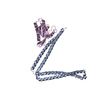
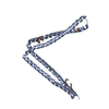
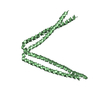
| Title | Crystal structure of SeMet apo SH3BP5 (I41) | ||||||
 Components Components | SH3 domain-binding protein 5 | ||||||
 Keywords Keywords |  SIGNALING PROTEIN / Rab11 / GEF / SH3BP5 SIGNALING PROTEIN / Rab11 / GEF / SH3BP5 | ||||||
| Function / homology |  Function and homology informationprotein kinase inhibitor activity / guanyl-nucleotide exchange factor activity / cytoplasmic vesicle membrane / SH3 domain binding / nuclear body / intracellular signal transduction / signal transduction / mitochondrion / nucleoplasm / cytoplasm Function and homology informationprotein kinase inhibitor activity / guanyl-nucleotide exchange factor activity / cytoplasmic vesicle membrane / SH3 domain binding / nuclear body / intracellular signal transduction / signal transduction / mitochondrion / nucleoplasm / cytoplasmSimilarity search - Function | ||||||
| Biological species |  Homo sapiens (human) Homo sapiens (human) | ||||||
| Method | X-RAY DIFFRACTION / SYNCHROTRON / SAD / Resolution: 3.35 Å | ||||||
 Authors Authors | Goto-Ito, S. / Yamagata, A. / Sato, Y. / Fukai, S. | ||||||
| Funding support |  Japan, 1items Japan, 1items
| ||||||
 Citation Citation | Journal: Life Sci Alliance / Year: 2019 Title: Structural basis of guanine nucleotide exchange for Rab11 by SH3BP5. Authors: Goto-Ito, S. / Morooka, N. / Yamagata, A. / Sato, Y. / Sato, K. / Fukai, S. | ||||||
| History |
|
- Structure visualization
| Structure viewer | Molecule: MolmilJmol/JSmol |
|---|
- Downloads & links
-Download
| PDBx/mmCIF format | 6ixe.cif.gz | 53.7 KB | Display | PDBx/mmCIF format |
|---|---|---|---|---|
| PDB format | pdb6ixe.ent.gz | 40.6 KB | Display | PDB format |
| PDBx/mmJSON format | 6ixe.json.gz | Tree view | PDBx/mmJSON format | |
| Others |  Other downloads Other downloads |
-Validation report
| Arichive directory | https://data.pdbj.org/pub/pdb/validation_reports/ix/6ixeftp://data.pdbj.org/pub/pdb/validation_reports/ix/6ixe | HTTPS FTP |
|---|
-Related structure data


-Links
 PDBj
PDBj- Assembly
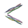
| Deposited unit | 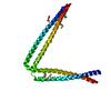
| ||||||||
|---|---|---|---|---|---|---|---|---|---|
| 1 |
| ||||||||
| Unit cell |
|
-Components
| #1: Protein | Mass: 26690.434 Da / Num. of mol.: 1 / Mutation: M167A,R260A,R261A,R262A Source method: isolated from a genetically manipulated source Source: (gene. exp.) Homo sapiens (human) / Gene: SH3BP5, SAB / Production host:  Escherichia coli (E. coli) / References: UniProt: O60239 Escherichia coli (E. coli) / References: UniProt: O60239 |
|---|---|
| #2: Chemical | ChemComp-SIN / Succinic acid  Mass: 118.088 Da / Num. of mol.: 1 / Source method: obtained synthetically / Formula: C4H6O4 Mass: 118.088 Da / Num. of mol.: 1 / Source method: obtained synthetically / Formula: C4H6O4 |
-Experimental details
-Experiment
| Experiment | Method: X-RAY DIFFRACTION / Number of used crystals: 1 |
|---|
- Sample preparation
| Crystal | Density Matthews: 3.18 Å3/Da / Density % sol: 61.29 % |
|---|---|
| Crystal grow | Temperature: 293 K / Method: vapor diffusion, sitting drop / Details: 0.1M succinic acid (pH 7.0), 12% PEG 3350 |
-Data collection
| Diffraction | Mean temperature: 100 K / Serial crystal experiment: N |
|---|---|
| Diffraction source | Source: SYNCHROTRON / Site: SPring-8 / Beamline: BL41XU / Wavelength: 0.9792 Å |
| Detector | Type: DECTRIS EIGER X 16M / Detector: PIXEL / Date: Jan 29, 2018 |
| Radiation | Protocol: SINGLE WAVELENGTH / Monochromatic (M) / Laue (L): M / Scattering type: x-ray |
| Radiation wavelength | Wavelength: 0.9792 Å / Relative weight: 1 |
| Reflection | Resolution: 3.35→50 Å / Num. obs: 4777 / % possible obs: 96.9 % / Redundancy: 8.2 % / Rsym value: 0.158 / Net I/σ(I): 30 |
| Reflection shell | Resolution: 3.35→3.41 Å / Rsym value: 0.357 |
- Processing
| Software |
| ||||||||||||||||||||||||
|---|---|---|---|---|---|---|---|---|---|---|---|---|---|---|---|---|---|---|---|---|---|---|---|---|---|
| Refinement | Method to determine structure: SAD / Resolution: 3.35→38.899 Å / SU ML: 0.65 / Cross valid method: FREE R-VALUE / σ(F): 1.6 / Phase error: 42.51 / Stereochemistry target values: ML
| ||||||||||||||||||||||||
| Solvent computation | Shrinkage radii: 0.9 Å / VDW probe radii: 1.11 Å / Solvent model: FLAT BULK SOLVENT MODEL | ||||||||||||||||||||||||
| Refinement step | Cycle: LAST / Resolution: 3.35→38.899 Å
| ||||||||||||||||||||||||
| Refine LS restraints |
| ||||||||||||||||||||||||
| LS refinement shell |
|
