 Movie
Movie Controller


+ Open data

- Basic information
| Entry | Database: PDB / ID: 6is6 | ||||||
|---|---|---|---|---|---|---|---|
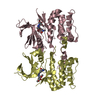
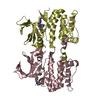
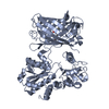
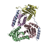
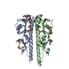
| Title | Crystal structure of Thermoplasmatales archaeon heliorhodopsin | ||||||
 Components Components | heliorhodopsin | ||||||
 Keywords Keywords | MEMBRANE PROTEIN / alpha-helical | ||||||
| Function / homology | Heliorhodopsin / Heliorhodopsin / membrane / identical protein binding / (2R)-2,3-dihydroxypropyl (9Z)-octadec-9-enoate / RETINAL / Heliorhodopsin HeR Function and homology information Function and homology information | ||||||
| Biological species |  Thermoplasmatales archaeon SG8-52-1 (archaea) Thermoplasmatales archaeon SG8-52-1 (archaea) | ||||||
| Method | X-RAY DIFFRACTION / SYNCHROTRON / MOLECULAR REPLACEMENT / Resolution: 2.4 Å | ||||||
 Authors Authors | Shihoya, W. / Yamashita, K. / Nureki, O. | ||||||
 Citation Citation | Journal: Nature / Year: 2019 Title: Crystal structure of heliorhodopsin. Authors: Shihoya, W. / Inoue, K. / Singh, M. / Konno, M. / Hososhima, S. / Yamashita, K. / Ikeda, K. / Higuchi, A. / Izume, T. / Okazaki, S. / Hashimoto, M. / Mizutori, R. / Tomida, S. / Yamauchi, Y. ...Authors: Shihoya, W. / Inoue, K. / Singh, M. / Konno, M. / Hososhima, S. / Yamashita, K. / Ikeda, K. / Higuchi, A. / Izume, T. / Okazaki, S. / Hashimoto, M. / Mizutori, R. / Tomida, S. / Yamauchi, Y. / Abe-Yoshizumi, R. / Katayama, K. / Tsunoda, S.P. / Shibata, M. / Furutani, Y. / Pushkarev, A. / Beja, O. / Uchihashi, T. / Kandori, H. / Nureki, O. | ||||||
| History |
|
- Structure visualization
| Structure viewer | Molecule: MolmilJmol/JSmol |
|---|
- Downloads & links
-Download
| PDBx/mmCIF format | 6is6.cif.gz | 76.6 KB | Display | PDBx/mmCIF format |
|---|---|---|---|---|
| PDB format | pdb6is6.ent.gz | 55.7 KB | Display | PDB format |
| PDBx/mmJSON format | 6is6.json.gz | Tree view | PDBx/mmJSON format | |
| Others |  Other downloads Other downloads |
-Validation report
| Arichive directory | https://data.pdbj.org/pub/pdb/validation_reports/is/6is6ftp://data.pdbj.org/pub/pdb/validation_reports/is/6is6 | HTTPS FTP |
|---|
-Related structure data
| Related structure data |  5ax0S S: Starting model for refinement |
|---|---|
| Similar structure data | |
| Experimental dataset #1 | Data reference: 10.5281/zenodo.3333323 / Data set type: diffraction image data / Metadata reference: 10.5281/zenodo.3333323 |


-Links
 PDBj
PDBj


- Assembly
| Deposited unit | 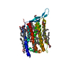
| ||||||||
|---|---|---|---|---|---|---|---|---|---|
| 1 | 
| ||||||||
| Unit cell |
|
-Components
| #1: Protein | Mass: 29886.301 Da / Num. of mol.: 1 Source method: isolated from a genetically manipulated source Source: (gene. exp.) Thermoplasmatales archaeon SG8-52-1 (archaea)Plasmid: pET21a(+) / Production host:  Escherichia coli (E. coli) / Variant (production host): C41(Rosetta) / References: UniProt: A0A151EDA9 Escherichia coli (E. coli) / Variant (production host): C41(Rosetta) / References: UniProt: A0A151EDA9 | ||||||
|---|---|---|---|---|---|---|---|
| #2: Chemical | ChemComp-OLC / (   Mass: 356.540 Da / Num. of mol.: 14 / Source method: obtained synthetically / Formula: C21H40O4 Mass: 356.540 Da / Num. of mol.: 14 / Source method: obtained synthetically / Formula: C21H40O4#3: Chemical | ChemComp-RET / | Retinal  Mass: 284.436 Da / Num. of mol.: 1 / Source method: isolated from a natural source / Formula: C20H28O / Feature type: SUBJECT OF INVESTIGATION Mass: 284.436 Da / Num. of mol.: 1 / Source method: isolated from a natural source / Formula: C20H28O / Feature type: SUBJECT OF INVESTIGATION#4: Water | ChemComp-HOH / | Water Mass: 18.015 Da / Num. of mol.: 31 / Source method: isolated from a natural source / Formula: H2O Mass: 18.015 Da / Num. of mol.: 31 / Source method: isolated from a natural source / Formula: H2OHas ligand of interest | Y | |
-Experimental details
-Experiment
| Experiment | Method: X-RAY DIFFRACTION / Number of used crystals: 1 |
|---|
- Sample preparation
| Crystal | Density Matthews: 2.57 Å3/Da / Density % sol: 52.06 % |
|---|---|
| Crystal grow | Temperature: 293 K / Method: lipidic cubic phase / pH: 8 Details: 30% PEG 350MME, 100mM Tris-HCl, pH 8.0, 100mM potassium citrate |
-Data collection
| Diffraction | Mean temperature: 100 K / Serial crystal experiment: N | ||||||||||||||||||||||||||||||||||||||||||||||||||||||||||||||||||||||||||||||||
|---|---|---|---|---|---|---|---|---|---|---|---|---|---|---|---|---|---|---|---|---|---|---|---|---|---|---|---|---|---|---|---|---|---|---|---|---|---|---|---|---|---|---|---|---|---|---|---|---|---|---|---|---|---|---|---|---|---|---|---|---|---|---|---|---|---|---|---|---|---|---|---|---|---|---|---|---|---|---|---|---|---|
| Diffraction source | Source: SYNCHROTRON / Site: SPring-8  / Beamline: BL32XU / Wavelength: 1 Å / Beamline: BL32XU / Wavelength: 1 Å | ||||||||||||||||||||||||||||||||||||||||||||||||||||||||||||||||||||||||||||||||
| Detector | Type: DECTRIS EIGER X 9M / Detector: PIXEL / Date: Apr 18, 2018 | ||||||||||||||||||||||||||||||||||||||||||||||||||||||||||||||||||||||||||||||||
| Radiation | Monochromator: Si(111) / Protocol: SINGLE WAVELENGTH / Monochromatic (M) / Laue (L): M / Scattering type: x-ray | ||||||||||||||||||||||||||||||||||||||||||||||||||||||||||||||||||||||||||||||||
| Radiation wavelength | Wavelength: 1 Å / Relative weight: 1 | ||||||||||||||||||||||||||||||||||||||||||||||||||||||||||||||||||||||||||||||||
| Reflection | Resolution: 2.4→48.773 Å / Num. obs: 12404 / % possible obs: 99.9 % / Redundancy: 35.302 % / Biso Wilson estimate: 32.63 Å2 / CC1/2: 0.995 / Rmerge(I) obs: 0.324 / Rrim(I) all: 0.328 / Χ2: 1.218 / Net I/σ(I): 13.68 / Num. measured all: 437886 / Scaling rejects: 318 | ||||||||||||||||||||||||||||||||||||||||||||||||||||||||||||||||||||||||||||||||
| Reflection shell | Diffraction-ID: 1
|
- Processing
| Software |
| ||||||||||||||||||||||||||||||
|---|---|---|---|---|---|---|---|---|---|---|---|---|---|---|---|---|---|---|---|---|---|---|---|---|---|---|---|---|---|---|---|
| Refinement | Method to determine structure: MOLECULAR REPLACEMENT Starting model: 5AX0 Resolution: 2.4→48.773 Å / SU ML: 0.25 / Cross valid method: THROUGHOUT / σ(F): 1.33 / Phase error: 24.09
| ||||||||||||||||||||||||||||||
| Solvent computation | Shrinkage radii: 0.9 Å / VDW probe radii: 1.11 Å | ||||||||||||||||||||||||||||||
| Displacement parameters | Biso max: 102.23 Å2 / Biso mean: 39.1079 Å2 / Biso min: 18.45 Å2 | ||||||||||||||||||||||||||||||
| Refinement step | Cycle: final / Resolution: 2.4→48.773 Å
| ||||||||||||||||||||||||||||||
| LS refinement shell | Refine-ID: X-RAY DIFFRACTION / Rfactor Rfree error: 0
|
