 Movie
Movie Controller

[English] 日本語
 Yorodumi
- PDB-6iqy: High resolution structure of bilirubin oxidase from Myrothecium v... -
+ Open data

- Basic information
| Entry | Database: PDB / ID: 6iqy | ||||||||||||
|---|---|---|---|---|---|---|---|---|---|---|---|---|---|
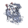
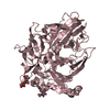
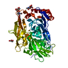
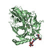
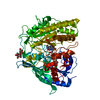
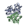
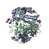
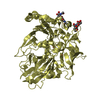
| Title | High resolution structure of bilirubin oxidase from Myrothecium verrucaria - M467Q mutant, anaerobically prepared | ||||||||||||
 Components Components | Bilirubin oxidase | ||||||||||||
 Keywords Keywords | OXIDOREDUCTASE / Multicopper oxydase | ||||||||||||
| Function / homology |  Function and homology information Function and homology information | ||||||||||||
| Biological species |  Myrothecium verrucaria (fungus) Myrothecium verrucaria (fungus) | ||||||||||||
| Method | X-RAY DIFFRACTION / SYNCHROTRON / MOLECULAR REPLACEMENT / Resolution: 1.6 Å | ||||||||||||
 Authors Authors | Shibata, N. / Akter, M. / Higuchi, Y. | ||||||||||||
 Citation Citation | Journal: Chemistry / Year: 2018 Title: Redox Potential-Dependent Formation of an Unusual His-Trp Bond in Bilirubin Oxidase. Authors: Akter, M. / Tokiwa, T. / Shoji, M. / Nishikawa, K. / Shigeta, Y. / Sakurai, T. / Higuchi, Y. / Kataoka, K. / Shibata, N. | ||||||||||||
| History |
|
- Structure visualization
| Structure viewer | Molecule: MolmilJmol/JSmol |
|---|
- Downloads & links
-Download
| PDBx/mmCIF format | 6iqy.cif.gz | 268.5 KB | Display | PDBx/mmCIF format |
|---|---|---|---|---|
| PDB format | pdb6iqy.ent.gz | 211.1 KB | Display | PDB format |
| PDBx/mmJSON format | 6iqy.json.gz | Tree view | PDBx/mmJSON format | |
| Others |  Other downloads Other downloads |
-Validation report
| Arichive directory | https://data.pdbj.org/pub/pdb/validation_reports/iq/6iqyftp://data.pdbj.org/pub/pdb/validation_reports/iq/6iqy | HTTPS FTP |
|---|
-Related structure data
| Related structure data |  6iqxC  6iqzC 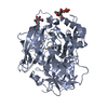 2xllS S: Starting model for refinement C: citing same article ( |
|---|---|
| Similar structure data |


-Links
 PDBj
PDBj- Assembly
| Deposited unit | 
| ||||||||
|---|---|---|---|---|---|---|---|---|---|
| 1 | 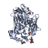
| ||||||||
| 2 | 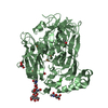
| ||||||||
| Unit cell |
|
-Components
-Protein , 1 types, 2 molecules AB
| #1: Protein | Mass: 60545.227 Da / Num. of mol.: 2 / Mutation: M467Q Source method: isolated from a genetically manipulated source Source: (gene. exp.) Myrothecium verrucaria (fungus) / Plasmid: pPICBO / Production host: Komagataella pastoris (fungus) / References: UniProt: Q12737, bilirubin oxidase |
|---|
-Sugars , 2 types, 4 molecules 
| #2: Polysaccharide | / Mass: 586.542 Da / Num. of mol.: 2 Source method: isolated from a genetically manipulated source #4: Sugar | N-Acetylglucosamine Type: D-saccharide, beta linking / Mass: 221.208 Da / Num. of mol.: 2 Type: D-saccharide, beta linking / Mass: 221.208 Da / Num. of mol.: 2Source method: isolated from a genetically manipulated source Formula: C8H15NO6 |
|---|
-Non-polymers , 5 types, 1500 molecules 


| #3: Chemical | ChemComp-CU / Copper Mass: 63.546 Da / Num. of mol.: 8 / Source method: obtained synthetically / Formula: Cu Mass: 63.546 Da / Num. of mol.: 8 / Source method: obtained synthetically / Formula: Cu#5: Chemical | Acetate Mass: 59.044 Da / Num. of mol.: 2 / Source method: obtained synthetically / Formula: C2H3O2 Mass: 59.044 Da / Num. of mol.: 2 / Source method: obtained synthetically / Formula: C2H3O2#6: Chemical | ChemComp-GOL / Glycerol Mass: 92.094 Da / Num. of mol.: 7 / Source method: obtained synthetically / Formula: C3H8O3 Mass: 92.094 Da / Num. of mol.: 7 / Source method: obtained synthetically / Formula: C3H8O3#7: Chemical |  Mass: 40.078 Da / Num. of mol.: 2 / Source method: obtained synthetically / Formula: Ca Mass: 40.078 Da / Num. of mol.: 2 / Source method: obtained synthetically / Formula: Ca#8: Water | ChemComp-HOH / | WaterMass: 18.015 Da / Num. of mol.: 1481 / Source method: isolated from a natural source / Formula: H2O |
|---|
-Experimental details
-Experiment
| Experiment | Method: X-RAY DIFFRACTION / Number of used crystals: 1 |
|---|
- Sample preparation
| Crystal | Density Matthews: 2.61 Å3/Da / Density % sol: 52.78 % |
|---|---|
| Crystal grow | Temperature: 293 K / Method: vapor diffusion, sitting drop Details: 0.1M MES PH 5.5, 12% (W/V) PEG 8000, 0.1M CALCIUM ACETATE, VAPOR DIFFUSION, SITTING DROP, TEMPERATURE 293K |
-Data collection
| Diffraction | Mean temperature: 100 K / Serial crystal experiment: N |
|---|---|
| Diffraction source | Source: SYNCHROTRON / Site: Photon Factory  / Beamline: BL-17A / Wavelength: 1 Å / Beamline: BL-17A / Wavelength: 1 Å |
| Detector | Type: DECTRIS PILATUS3 S 6M / Detector: PIXEL / Date: Mar 4, 2016 |
| Radiation | Protocol: SINGLE WAVELENGTH / Monochromatic (M) / Laue (L): M / Scattering type: x-ray |
| Radiation wavelength | Wavelength: 1 Å / Relative weight: 1 |
| Reflection | Resolution: 1.6→50 Å / Num. obs: 162206 / % possible obs: 99.7 % / Redundancy: 3.3 % / Rmerge(I) obs: 0.125 / Net I/σ(I): 11.49 |
| Reflection shell | Resolution: 1.6→1.63 Å / Redundancy: 3.3 % / Rmerge(I) obs: 0.557 / Mean I/σ(I) obs: 2.22 / % possible all: 99.7 |
- Processing
| Software |
| |||||||||||||||||||||||||||||||||||||||||||||||||||||||||||||||||||||||||||||||||||||||||||||||||||||||||
|---|---|---|---|---|---|---|---|---|---|---|---|---|---|---|---|---|---|---|---|---|---|---|---|---|---|---|---|---|---|---|---|---|---|---|---|---|---|---|---|---|---|---|---|---|---|---|---|---|---|---|---|---|---|---|---|---|---|---|---|---|---|---|---|---|---|---|---|---|---|---|---|---|---|---|---|---|---|---|---|---|---|---|---|---|---|---|---|---|---|---|---|---|---|---|---|---|---|---|---|---|---|---|---|---|---|---|
| Refinement | Method to determine structure: MOLECULAR REPLACEMENT Starting model: 2XLL Resolution: 1.6→45.861 Å / SU ML: 0.2 / Cross valid method: FREE R-VALUE / σ(F): 1.35 / Phase error: 24.08
| |||||||||||||||||||||||||||||||||||||||||||||||||||||||||||||||||||||||||||||||||||||||||||||||||||||||||
| Solvent computation | Shrinkage radii: 0.9 Å / VDW probe radii: 1.11 Å | |||||||||||||||||||||||||||||||||||||||||||||||||||||||||||||||||||||||||||||||||||||||||||||||||||||||||
| Refinement step | Cycle: LAST / Resolution: 1.6→45.861 Å
| |||||||||||||||||||||||||||||||||||||||||||||||||||||||||||||||||||||||||||||||||||||||||||||||||||||||||
| Refine LS restraints |
| |||||||||||||||||||||||||||||||||||||||||||||||||||||||||||||||||||||||||||||||||||||||||||||||||||||||||
| LS refinement shell |
|
