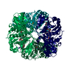
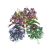
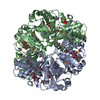
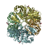
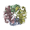
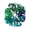
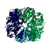
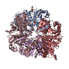
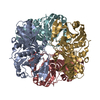
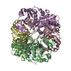
Entry Database : PDB / ID : 6iq6Title Crystal structure of GAPDH Glyceraldehyde-3-phosphate dehydrogenase Keywords / / / Function / homology Function Domain/homology Component
/ / / / / / / / / / / / / / / / / / / / / / / / / / / / / / / / / / / / / / / / / / / / / / / / / / / / / / / / / / / / / / / / / / / / / / / / / Biological species Homo sapiens (human)Method / / / Resolution : 2.29 Å Authors Park, J.B. / Park, H.Y. Journal : Mol.Cells / Year : 2019Title : Structural Study of Monomethyl Fumarate-Bound Human GAPDH.Authors : Park, J.B. / Park, H. / Son, J. / Ha, S.J. / Cho, H.S. History Deposition Nov 6, 2018 Deposition site / Processing site Revision 1.0 Aug 28, 2019 Provider / Type Revision 1.1 Sep 11, 2019 Group / Database references / Category Item / _citation.page_first / _citation.page_lastRevision 1.2 Nov 22, 2023 Group / Database references / Refinement descriptionCategory chem_comp_atom / chem_comp_bond ... chem_comp_atom / chem_comp_bond / database_2 / pdbx_initial_refinement_model Item / _database_2.pdbx_database_accession
Show all Show less
 Movie
Movie Controller


 Open data
 Basic information
 Components
 Glyceraldehyde 3-phosphate dehydrogenase
  Keywords
 Function and homology information
 Authors
 Citation
 Structure visualization
 Downloads & links
 Other downloads
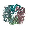


 PDBj
PDBj

 Assembly


 Mass: 130.099 Da / Num. of mol.: 5 / Source method: obtained synthetically / Formula: C5H6O4 / Feature type: SUBJECT OF INVESTIGATION
 Mass: 18.015 Da / Num. of mol.: 69 / Source method: isolated from a natural source / Formula: H2O
 Sample preparation
 / Beamline: 7A (6B, 6C1) / Wavelength: 0.987 Å
 Processing