 Movie
Movie Controller

[English] 日本語
 Yorodumi
- PDB-6inz: Crystal structure of solute-binding protein complexed with unsatu... -
+ Open data

- Basic information
| Entry | Database: PDB / ID: 6inz | |||||||||
|---|---|---|---|---|---|---|---|---|---|---|
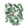
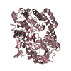
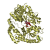
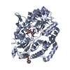
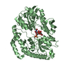
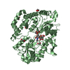
| Title | Crystal structure of solute-binding protein complexed with unsaturated hyaluronan disaccharide | |||||||||
 Components Components | Extracellular solute-binding protein family 1 | |||||||||
 Keywords Keywords | SUGAR BINDING PROTEIN /  glycosaminoglycan / ABC transporter / solute-binding protein glycosaminoglycan / ABC transporter / solute-binding protein | |||||||||
| Function / homology | Bacterial extracellular solute-binding protein / Bacterial extracellular solute-binding protein / Periplasmic binding protein-like II / D-Maltodextrin-Binding Protein; domain 2 / Prokaryotic membrane lipoprotein lipid attachment site profile. / 3-Layer(aba) Sandwich / metal ion binding / Alpha Beta / Extracellular solute-binding protein family 1 Function and homology information Function and homology information | |||||||||
| Biological species |  Streptobacillus moniliformis DSM 12112 (bacteria) Streptobacillus moniliformis DSM 12112 (bacteria) | |||||||||
| Method | X-RAY DIFFRACTION / SYNCHROTRON / MOLECULAR REPLACEMENT / Resolution: 2.289 Å | |||||||||
 Authors Authors | Oiki, S. / Mikami, B. / Murata, K. / Hashimoto, W. | |||||||||
| Funding support |  Japan, 1items Japan, 1items
| |||||||||
 Citation Citation | Journal: Biosci.Biotechnol.Biochem. / Year: 2019 Title: Substrate recognition by bacterial solute-binding protein is responsible for import of extracellular hyaluronan and chondroitin sulfate from the animal host. Authors: Oiki, S. / Sato, M. / Mikami, B. / Murata, K. / Hashimoto, W. | |||||||||
| History |
|
- Structure visualization
| Structure viewer | Molecule: MolmilJmol/JSmol |
|---|
- Downloads & links
-Download
| PDBx/mmCIF format | 6inz.cif.gz | 121.3 KB | Display | PDBx/mmCIF format |
|---|---|---|---|---|
| PDB format | pdb6inz.ent.gz | 89.5 KB | Display | PDB format |
| PDBx/mmJSON format | 6inz.json.gz | Tree view | PDBx/mmJSON format | |
| Others |  Other downloads Other downloads |
-Validation report
| Arichive directory | https://data.pdbj.org/pub/pdb/validation_reports/in/6inzftp://data.pdbj.org/pub/pdb/validation_reports/in/6inz | HTTPS FTP |
|---|
-Related structure data
| Related structure data |  5gubS S: Starting model for refinement |
|---|---|
| Similar structure data |


-Links
 PDBj
PDBj


- Assembly
| Deposited unit | 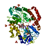
| ||||||||
|---|---|---|---|---|---|---|---|---|---|
| 1 |
| ||||||||
| Unit cell |
|
-Components
| #1: Protein | Mass: 57532.223 Da / Num. of mol.: 1 Source method: isolated from a genetically manipulated source Source: (gene. exp.) Streptobacillus moniliformis DSM 12112 (bacteria)Strain: DSM 12112 / Gene: Smon_0123 / Production host: Escherichia coli (E. coli) / References: UniProt: D1AWE0 | ||||
|---|---|---|---|---|---|
| #2: Polysaccharide | 4-deoxy-alpha-L-threo-hex-4-enopyranuronic acid-(1-3)-2-acetamido-2-deoxy-beta-D-glucopyranose / Mass: 379.317 Da / Num. of mol.: 1 Source method: isolated from a genetically manipulated source | ||||
| #3: Chemical | ChemComp-SO4 / Sulfate  Mass: 96.063 Da / Num. of mol.: 8 Mass: 96.063 Da / Num. of mol.: 8Source method: isolated from a genetically manipulated source Formula: SO4 #4: Chemical | ChemComp-CA / |   Mass: 40.078 Da / Num. of mol.: 1 Mass: 40.078 Da / Num. of mol.: 1Source method: isolated from a genetically manipulated source Formula: Ca #5: Water | ChemComp-HOH / | Water Mass: 18.015 Da / Num. of mol.: 272 / Source method: isolated from a natural source / Formula: H2O Mass: 18.015 Da / Num. of mol.: 272 / Source method: isolated from a natural source / Formula: H2O |
-Experimental details
-Experiment
| Experiment | Method: X-RAY DIFFRACTION / Number of used crystals: 1 |
|---|
- Sample preparation
| Crystal | Density Matthews: 2.73 Å3/Da / Density % sol: 55.03 % |
|---|---|
| Crystal grow | Temperature: 293 K / Method: vapor diffusion, sitting drop Details: 200mM litium sulfate, 100mM Tris-HCl (pH 8.5), 30% PEG 4000 |
-Data collection
| Diffraction | Mean temperature: 100 K / Serial crystal experiment: N |
|---|---|
| Diffraction source | Source: SYNCHROTRON / Site: SPring-8 / Beamline: BL26B1 / Wavelength: 1 Å |
| Detector | Type: RAYONIX MX225HE / Detector: CCD / Date: Jul 28, 2018 |
| Radiation | Protocol: SINGLE WAVELENGTH / Monochromatic (M) / Laue (L): M / Scattering type: x-ray |
| Radiation wavelength | Wavelength: 1 Å / Relative weight: 1 |
| Reflection | Resolution: 2.289→50 Å / Num. obs: 29129 / % possible obs: 99.9 % / Redundancy: 3.1 % / Rmerge(I) obs: 0.076 / Net I/σ(I): 23.6 |
| Reflection shell | Resolution: 2.289→2.34 Å / Rmerge(I) obs: 0.51 / Num. unique obs: 1439 |
- Processing
| Software |
| |||||||||||||||||||||||||||||||||||||||||||||||||||||||||||||||||||||||||||||
|---|---|---|---|---|---|---|---|---|---|---|---|---|---|---|---|---|---|---|---|---|---|---|---|---|---|---|---|---|---|---|---|---|---|---|---|---|---|---|---|---|---|---|---|---|---|---|---|---|---|---|---|---|---|---|---|---|---|---|---|---|---|---|---|---|---|---|---|---|---|---|---|---|---|---|---|---|---|---|
| Refinement | Method to determine structure: MOLECULAR REPLACEMENT Starting model: 5GUB Resolution: 2.289→41.41 Å / SU ML: 0.23 / Cross valid method: FREE R-VALUE / σ(F): 1.34 / Phase error: 21.81
| |||||||||||||||||||||||||||||||||||||||||||||||||||||||||||||||||||||||||||||
| Solvent computation | Shrinkage radii: 0.9 Å / VDW probe radii: 1.11 Å | |||||||||||||||||||||||||||||||||||||||||||||||||||||||||||||||||||||||||||||
| Refinement step | Cycle: LAST / Resolution: 2.289→41.41 Å
| |||||||||||||||||||||||||||||||||||||||||||||||||||||||||||||||||||||||||||||
| Refine LS restraints |
| |||||||||||||||||||||||||||||||||||||||||||||||||||||||||||||||||||||||||||||
| LS refinement shell |
|
