 Movie
Movie Controller


+ Open data

- Basic information
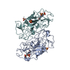
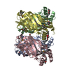
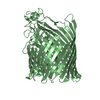
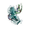
| Entry | Database: PDB / ID: 6iml | ||||||
|---|---|---|---|---|---|---|---|
| Title | The crystal structure of AsfvLIG:CT1 complex | ||||||
 Components Components |
| ||||||
 Keywords Keywords | LIGASE/DNA / The crysatl structure of AsfvLIG with C:G complex / LIGASE-DNA complex /  DNA BINDING PROTEIN DNA BINDING PROTEIN | ||||||
| Function / homology |  Function and homology informationDNA ligase (ATP) activity / DNA recombination / DNA replication / DNA repair / ATP binding Function and homology informationDNA ligase (ATP) activity / DNA recombination / DNA replication / DNA repair / ATP bindingSimilarity search - Function | ||||||
| Biological species |   African swine fever virus African swine fever virussynthetic construct (others) | ||||||
| Method | X-RAY DIFFRACTION / SYNCHROTRON / MOLECULAR REPLACEMENT / Resolution: 2.35 Å | ||||||
 Authors Authors | Chen, Y.Q. / Gan, J.H. | ||||||
| Funding support |  China, 1items China, 1items
| ||||||
 Citation Citation | Journal: Nat Commun / Year: 2019 Title: Structure of the error-prone DNA ligase of African swine fever virus identifies critical active site residues. Authors: Chen, Y. / Liu, H. / Yang, C. / Gao, Y. / Yu, X. / Chen, X. / Cui, R. / Zheng, L. / Li, S. / Li, X. / Ma, J. / Huang, Z. / Li, J. / Gan, J. | ||||||
| History |
|
- Structure visualization
| Structure viewer | Molecule: MolmilJmol/JSmol |
|---|
- Downloads & links
-Download
| PDBx/mmCIF format | 6iml.cif.gz | 121.1 KB | Display | PDBx/mmCIF format |
|---|---|---|---|---|
| PDB format | pdb6iml.ent.gz | 87.5 KB | Display | PDB format |
| PDBx/mmJSON format | 6iml.json.gz | Tree view | PDBx/mmJSON format | |
| Others |  Other downloads Other downloads |
-Validation report
| Arichive directory | https://data.pdbj.org/pub/pdb/validation_reports/im/6imlftp://data.pdbj.org/pub/pdb/validation_reports/im/6iml | HTTPS FTP |
|---|
-Related structure data
| Related structure data |  6imjC  6imkC  6imnC 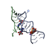 6ymjS S: Starting model for refinement C: citing same article ( |
|---|---|
| Similar structure data |


-Links
 PDBj
PDBj


- Assembly
| Deposited unit | 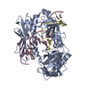
| ||||||||
|---|---|---|---|---|---|---|---|---|---|
| 1 |
| ||||||||
| Unit cell |
|
-Components
| #1: Protein | / NP419L / PNP419L Mass: 48233.562 Da / Num. of mol.: 1 Source method: isolated from a genetically manipulated source Source: (gene. exp.) African swine fever virus / Gene: NP419L / Production host:  Escherichia coli BL21(DE3) (bacteria) / Strain (production host): BL21(DE3) / References: UniProt: A0A0A1E0U0 Escherichia coli BL21(DE3) (bacteria) / Strain (production host): BL21(DE3) / References: UniProt: A0A0A1E0U0 |
|---|---|
| #2: DNA chain | Mass: 6643.284 Da / Num. of mol.: 1 / Source method: obtained synthetically / Source: (synth.) synthetic construct (others) |
| #3: DNA chain | Mass: 3694.402 Da / Num. of mol.: 1 / Source method: obtained synthetically / Source: (synth.) synthetic construct (others) |
| #4: DNA chain | Mass: 3101.028 Da / Num. of mol.: 1 / Source method: obtained synthetically / Source: (synth.) synthetic construct (others) |
| #5: Water | ChemComp-HOH / Water Mass: 18.015 Da / Num. of mol.: 67 / Source method: isolated from a natural source / Formula: H2O Mass: 18.015 Da / Num. of mol.: 67 / Source method: isolated from a natural source / Formula: H2O |
-Experimental details
-Experiment
| Experiment | Method: X-RAY DIFFRACTION / Number of used crystals: 1 |
|---|
- Sample preparation
| Crystal | Density Matthews: 2.36 Å3/Da / Density % sol: 47.87 % |
|---|---|
| Crystal grow | Temperature: 291 K / Method: vapor diffusion, hanging drop / pH: 5.5 Details: 0.1 M BIS-TRIS pH 5.5, 15% w/v PEG 10000, 0.1 M Ammonium acetate |
-Data collection
| Diffraction | Mean temperature: 100 K / Serial crystal experiment: N |
|---|---|
| Diffraction source | Source: SYNCHROTRON / Site: SSRF / Beamline: BL19U1 / Wavelength: 1 Å |
| Detector | Type: MARMOSAIC 300 mm CCD / Detector: CCD / Date: Jun 21, 2015 |
| Radiation | Protocol: SINGLE WAVELENGTH / Monochromatic (M) / Laue (L): M / Scattering type: x-ray |
| Radiation wavelength | Wavelength: 1 Å / Relative weight: 1 |
| Reflection | Resolution: 2.35→30 Å / Num. obs: 23122 / % possible obs: 95.5 % / Redundancy: 3.9 % / Rmerge(I) obs: 0.133 / Net I/σ(I): 15.9 |
| Reflection shell | Resolution: 2.35→2.43 Å / Redundancy: 2.5 % / Rmerge(I) obs: 0.6 / Mean I/σ(I) obs: 1.77 / Num. unique obs: 2021 / % possible all: 83.7 |
- Processing
| Software |
| |||||||||||||||||||||||||||||||||||||||||||||||||||||||||||||||
|---|---|---|---|---|---|---|---|---|---|---|---|---|---|---|---|---|---|---|---|---|---|---|---|---|---|---|---|---|---|---|---|---|---|---|---|---|---|---|---|---|---|---|---|---|---|---|---|---|---|---|---|---|---|---|---|---|---|---|---|---|---|---|---|---|
| Refinement | Method to determine structure: MOLECULAR REPLACEMENT Starting model: 6YMJ Resolution: 2.35→29.884 Å / SU ML: 0.33 / Cross valid method: FREE R-VALUE / σ(F): 1.35 / Phase error: 30.65
| |||||||||||||||||||||||||||||||||||||||||||||||||||||||||||||||
| Solvent computation | Shrinkage radii: 0.9 Å / VDW probe radii: 1.11 Å | |||||||||||||||||||||||||||||||||||||||||||||||||||||||||||||||
| Refinement step | Cycle: LAST / Resolution: 2.35→29.884 Å
| |||||||||||||||||||||||||||||||||||||||||||||||||||||||||||||||
| Refine LS restraints |
| |||||||||||||||||||||||||||||||||||||||||||||||||||||||||||||||
| LS refinement shell |
|
