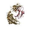
- PDB-6ilg: CRYSTAL STRUCTURE OF BAT MHC CLASS I PTAL-N*01:01 FOR 2.6 ANGSTROM -
+
Open data
ID or keywords:
Loading...
-
Basic information
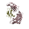
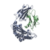
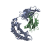
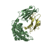
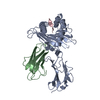
Entry
Database: PDB / ID: 6ilg
Title
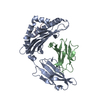
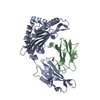
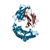
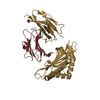
CRYSTAL STRUCTURE OF BAT MHC CLASS I PTAL-N*01:01 FOR 2.6 ANGSTROM
Components
Beta-2-microglobulinBeta-2 microglobulin
HEV-1-P8L
MHC class I antigen
Keywords
IMMUNE SYSTEM / IMMUNOLOGY / VIRUS
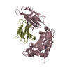
Function / homology
Function and homology information
antigen processing and presentation of peptide antigen via MHC class I / antigen processing and presentation of endogenous peptide antigen via MHC class I via ER pathway, TAP-independent / antigen processing and presentation of endogenous peptide antigen via MHC class Ib / lumenal side of endoplasmic reticulum membrane / MHC class I protein complex / positive regulation of T cell mediated cytotoxicity / phagocytic vesicle membrane / peptide antigen binding / disordered domain specific binding / amyloid fibril formation ...antigen processing and presentation of peptide antigen via MHC class I / antigen processing and presentation of endogenous peptide antigen via MHC class I via ER pathway, TAP-independent / antigen processing and presentation of endogenous peptide antigen via MHC class Ib / lumenal side of endoplasmic reticulum membrane / MHC class I protein complex / positive regulation of T cell mediated cytotoxicity / phagocytic vesicle membrane / peptide antigen binding / disordered domain specific binding / amyloid fibril formation / immune response / external side of plasma membrane / signaling receptor binding / protein-containing complex / extracellular space / extracellular region Similarity search - Function
Phosphoprotein P region PNT disordered / Phosphoprotein P region PNT disordered / Paramyxovirus structural protein P/V, N-terminal domain / Paramyxovirus structural protein V/P N-terminus / Phosphoprotein P soyouz module / N-terminal region of Paramyxovirinae phosphoprotein (P) / P/V phosphoprotein, paramyxoviral / Paramyxovirus P/V phosphoprotein C-terminal / MHC class I, alpha chain, C-terminal / MHC_I C-terminus ...Phosphoprotein P region PNT disordered / Phosphoprotein P region PNT disordered / Paramyxovirus structural protein P/V, N-terminal domain / Paramyxovirus structural protein V/P N-terminus / Phosphoprotein P soyouz module / N-terminal region of Paramyxovirinae phosphoprotein (P) / P/V phosphoprotein, paramyxoviral / Paramyxovirus P/V phosphoprotein C-terminal / MHC class I, alpha chain, C-terminal / MHC_I C-terminus / MHC class I-like antigen recognition-like / Murine Class I Major Histocompatibility Complex, H2-DB; Chain A, domain 1 / MHC class I alpha chain, alpha1 alpha2 domains / Class I Histocompatibility antigen, domains alpha 1 and 2 / Beta-2-Microglobulin / MHC class I-like antigen recognition-like / MHC class I-like antigen recognition-like superfamily / MHC classes I/II-like antigen recognition protein / Immunoglobulin/major histocompatibility complex, conserved site / Immunoglobulins and major histocompatibility complex proteins signature. / Immunoglobulin C-Type / Immunoglobulin C1-set / Immunoglobulin C1-set domain / Ig-like domain profile. / Immunoglobulin-like domain / Immunoglobulin-like domain superfamily / Nucleic acid-binding, OB-fold / Immunoglobulins / Immunoglobulin-like fold / Immunoglobulin-like / Sandwich / 2-Layer Sandwich / Mainly Beta / Alpha Beta Similarity search - Domain/homology
 Movie
Movie Controller

 Yorodumi
 Open data
 Basic information
 Components
 Keywords
 IMMUNE SYSTEM /
  Function and homology information
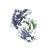
 Hendra virus
 Authors
 Citation
 Structure visualization
 Downloads & links
 Other downloads


 PDBj
PDBj


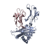
 Assembly

 Mass: 18.015 Da / Num. of mol.: 116 / Source method: isolated from a natural source / Formula: H2O
 Sample preparation
 Processing