 Movie
Movie Controller

[English] 日本語
 Yorodumi
- PDB-6ijf: Crystal structure of the type VI effector-immunity complex (Tae4-... -
+ Open data

- Basic information
| Entry | Database: PDB / ID: 6ijf | ||||||
|---|---|---|---|---|---|---|---|
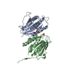
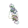
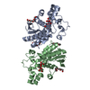
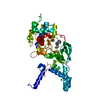
| Title | Crystal structure of the type VI effector-immunity complex (Tae4-Tai4) from Agrobacterium tumefaciens | ||||||
 Components Components |
| ||||||
 Keywords Keywords |  ANTITOXIN / COMPLEX ANTITOXIN / COMPLEX | ||||||
| Function / homology |  Function and homology information Function and homology informationendopeptidase fold (from Nostoc punctiforme) - #80 / MYOD Basic-Helix-Loop-Helix Domain, subunit B - #80 / Rap1a immunity protein / Rap1a immunity proteins / Type VI secretion system (T6SS), amidase effector protein 4 / Type VI secretion system (T6SS), amidase effector protein 4 / MYOD Basic-Helix-Loop-Helix Domain, subunit B / endopeptidase fold (from Nostoc punctiforme) / Few Secondary Structures / Irregular ...endopeptidase fold (from Nostoc punctiforme) - #80 / MYOD Basic-Helix-Loop-Helix Domain, subunit B - #80 / Rap1a immunity protein / Rap1a immunity proteins / Type VI secretion system (T6SS), amidase effector protein 4 / Type VI secretion system (T6SS), amidase effector protein 4 / MYOD Basic-Helix-Loop-Helix Domain, subunit B / endopeptidase fold (from Nostoc punctiforme) / Few Secondary Structures / Irregular / Alpha-Beta Complex / Alpha BetaSimilarity search - Domain/homology | ||||||
| Biological species |  Agrobacterium tumefaciens (bacteria) Agrobacterium tumefaciens (bacteria) | ||||||
| Method | X-RAY DIFFRACTION / SYNCHROTRON / MOLECULAR REPLACEMENT / Resolution: 1.9 Å | ||||||
 Authors Authors | Fukuhara, S. / Nakane, T. / Yamashita, K. / Ishii, R. / Ishitani, R. / Nureki, O. | ||||||
 Citation Citation | Journal: Acta Crystallogr F Struct Biol Commun / Year: 2018 Title: Crystal structure of the Agrobacterium tumefaciens type VI effector-immunity complex. Authors: Fukuhara, S. / Nakane, T. / Yamashita, K. / Ishii, R. / Ishitani, R. / Nureki, O. | ||||||
| History |
|
- Structure visualization
| Structure viewer | Molecule: MolmilJmol/JSmol |
|---|
- Downloads & links
-Download
| PDBx/mmCIF format | 6ijf.cif.gz | 118.1 KB | Display | PDBx/mmCIF format |
|---|---|---|---|---|
| PDB format | pdb6ijf.ent.gz | 89.7 KB | Display | PDB format |
| PDBx/mmJSON format | 6ijf.json.gz | Tree view | PDBx/mmJSON format | |
| Others |  Other downloads Other downloads |
-Validation report
| Arichive directory | https://data.pdbj.org/pub/pdb/validation_reports/ij/6ijfftp://data.pdbj.org/pub/pdb/validation_reports/ij/6ijf | HTTPS FTP |
|---|
-Related structure data
| Related structure data |  6ijeC  4bi8S S: Starting model for refinement C: citing same article ( |
|---|---|
| Similar structure data | |
| Experimental dataset #1 | Data reference: 10.5281/zenodo.1453302 / Data set type: diffraction image data |


-Links
 PDBj
PDBj- Assembly
| Deposited unit | 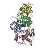
| ||||||||||||||||||||||||||||||||||||||||||||||||||||||||||||||||||||
|---|---|---|---|---|---|---|---|---|---|---|---|---|---|---|---|---|---|---|---|---|---|---|---|---|---|---|---|---|---|---|---|---|---|---|---|---|---|---|---|---|---|---|---|---|---|---|---|---|---|---|---|---|---|---|---|---|---|---|---|---|---|---|---|---|---|---|---|---|---|
| 1 |
| ||||||||||||||||||||||||||||||||||||||||||||||||||||||||||||||||||||
| Unit cell |
| ||||||||||||||||||||||||||||||||||||||||||||||||||||||||||||||||||||
| Noncrystallographic symmetry (NCS) | NCS domain:
NCS domain segments: Component-ID: 0 / Refine code: 0
NCS ensembles :
|
-Components
| #1: Protein | Mass: 11482.844 Da / Num. of mol.: 2 Source method: isolated from a genetically manipulated source Source: (gene. exp.) Agrobacterium tumefaciens (bacteria) / Gene: SY94_4314 / Plasmid: modified pETDuet-1 / Production host: Escherichia coli (E. coli) / Strain (production host): Rosetta 2 (DE3) / References: UniProt: A0A083ZID3#2: Protein | Mass: 18120.299 Da / Num. of mol.: 2 Source method: isolated from a genetically manipulated source Source: (gene. exp.) Agrobacterium tumefaciens (bacteria) / Gene: SY94_4315 / Plasmid: modified pETDuet-1 / Production host: Escherichia coli (E. coli) / Strain (production host): Rosetta 2 (DE3) / References: UniProt: A0A083ZID4#3: Chemical | ChemComp-1PE / | Polyethylene glycol  Mass: 238.278 Da / Num. of mol.: 1 / Source method: obtained synthetically / Formula: C10H22O6 / Comment: precipitant*YM Mass: 238.278 Da / Num. of mol.: 1 / Source method: obtained synthetically / Formula: C10H22O6 / Comment: precipitant*YM#4: Chemical | ChemComp-SO4 / | Sulfate  Mass: 96.063 Da / Num. of mol.: 1 / Source method: obtained synthetically / Formula: SO4 Mass: 96.063 Da / Num. of mol.: 1 / Source method: obtained synthetically / Formula: SO4#5: Water | ChemComp-HOH / | Water Mass: 18.015 Da / Num. of mol.: 122 / Source method: isolated from a natural source / Formula: H2O Mass: 18.015 Da / Num. of mol.: 122 / Source method: isolated from a natural source / Formula: H2O |
|---|
-Experimental details
-Experiment
| Experiment | Method: X-RAY DIFFRACTION / Number of used crystals: 1 |
|---|
- Sample preparation
| Crystal | Density Matthews: 2.46 Å3/Da / Density % sol: 49.96 % / Mosaicity: 0 ° |
|---|---|
| Crystal grow | Temperature: 293 K / Method: vapor diffusion, sitting drop / pH: 8.5 Details: 35 % PEG 400, 0.05 M Tris pH 8.5, 0.05 M sodium sulfate, 0.05 M lithium sulfate |
-Data collection
| Diffraction | Mean temperature: 100 K / Serial crystal experiment: N | ||||||||||||||||||||||||
|---|---|---|---|---|---|---|---|---|---|---|---|---|---|---|---|---|---|---|---|---|---|---|---|---|---|
| Diffraction source | Source: SYNCHROTRON / Site: SPring-8  / Beamline: BL32XU / Wavelength: 1 Å / Beamline: BL32XU / Wavelength: 1 Å | ||||||||||||||||||||||||
| Detector | Type: RAYONIX MX225-HS / Detector: CCD / Date: Dec 16, 2015 | ||||||||||||||||||||||||
| Radiation | Monochromator: Si(111) / Protocol: SINGLE WAVELENGTH / Monochromatic (M) / Laue (L): M / Scattering type: x-ray | ||||||||||||||||||||||||
| Radiation wavelength | Wavelength: 1 Å / Relative weight: 1 | ||||||||||||||||||||||||
| Reflection | Resolution: 1.9→97.18 Å / Num. obs: 44633 / % possible obs: 99.6 % / Redundancy: 10.1 % / CC1/2: 0.999 / Rmerge(I) obs: 0.067 / Rpim(I) all: 0.022 / Rrim(I) all: 0.07 / Net I/σ(I): 15 / Num. measured all: 452487 / Scaling rejects: 117 | ||||||||||||||||||||||||
| Reflection shell | Diffraction-ID: 1
|
- Processing
| Software |
| ||||||||||||||||||||||||||||||||||||||||||||||||||||||||||||||||||||||||||||||||||||||||||||||||||||||||||||||||||||||||||||||||||||||||||||||||||||||||||||||||||||||||||||||||||||||
|---|---|---|---|---|---|---|---|---|---|---|---|---|---|---|---|---|---|---|---|---|---|---|---|---|---|---|---|---|---|---|---|---|---|---|---|---|---|---|---|---|---|---|---|---|---|---|---|---|---|---|---|---|---|---|---|---|---|---|---|---|---|---|---|---|---|---|---|---|---|---|---|---|---|---|---|---|---|---|---|---|---|---|---|---|---|---|---|---|---|---|---|---|---|---|---|---|---|---|---|---|---|---|---|---|---|---|---|---|---|---|---|---|---|---|---|---|---|---|---|---|---|---|---|---|---|---|---|---|---|---|---|---|---|---|---|---|---|---|---|---|---|---|---|---|---|---|---|---|---|---|---|---|---|---|---|---|---|---|---|---|---|---|---|---|---|---|---|---|---|---|---|---|---|---|---|---|---|---|---|---|---|---|---|
| Refinement | Method to determine structure: MOLECULAR REPLACEMENT Starting model: 4BI8 Resolution: 1.9→62.38 Å / Cor.coef. Fo:Fc: 0.971 / Cor.coef. Fo:Fc free: 0.964 / SU B: 4.379 / SU ML: 0.121 / Cross valid method: THROUGHOUT / ESU R: 0.149 / ESU R Free: 0.13 / Details: HYDROGENS HAVE BEEN ADDED IN THE RIDING POSITIONS
| ||||||||||||||||||||||||||||||||||||||||||||||||||||||||||||||||||||||||||||||||||||||||||||||||||||||||||||||||||||||||||||||||||||||||||||||||||||||||||||||||||||||||||||||||||||||
| Solvent computation | Ion probe radii: 0.8 Å / Shrinkage radii: 0.8 Å / VDW probe radii: 1.2 Å | ||||||||||||||||||||||||||||||||||||||||||||||||||||||||||||||||||||||||||||||||||||||||||||||||||||||||||||||||||||||||||||||||||||||||||||||||||||||||||||||||||||||||||||||||||||||
| Displacement parameters | Biso mean: 47.589 Å2
| ||||||||||||||||||||||||||||||||||||||||||||||||||||||||||||||||||||||||||||||||||||||||||||||||||||||||||||||||||||||||||||||||||||||||||||||||||||||||||||||||||||||||||||||||||||||
| Refinement step | Cycle: 1 / Resolution: 1.9→62.38 Å
| ||||||||||||||||||||||||||||||||||||||||||||||||||||||||||||||||||||||||||||||||||||||||||||||||||||||||||||||||||||||||||||||||||||||||||||||||||||||||||||||||||||||||||||||||||||||
| Refine LS restraints |
|