 Movie
Movie Controller


+ Open data

- Basic information
| Entry | Database: PDB / ID: 6iev | |||||||||
|---|---|---|---|---|---|---|---|---|---|---|
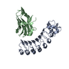
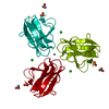
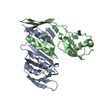
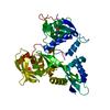
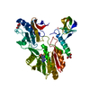
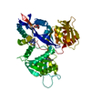
| Title | Crystal structure of a designed protein | |||||||||
 Components Components | Designed protein Design Design | |||||||||
 Keywords Keywords | STRUCTURAL PROTEIN / Thioredoxin / Enzyme / Folded / designed | |||||||||
| Function / homology | Glutaredoxin / Glutaredoxin / 3-Layer(aba) Sandwich / Alpha Beta Function and homology information Function and homology information | |||||||||
| Biological species |  Trypanosoma brucei (eukaryote) Trypanosoma brucei (eukaryote) | |||||||||
| Method | X-RAY DIFFRACTION / SYNCHROTRON / MOLECULAR REPLACEMENT / Resolution: 2.25 Å | |||||||||
 Authors Authors | Han, M. / Liao, S. / Chen, Q. / Liu, H. | |||||||||
| Funding support |  China, 2items China, 2items
| |||||||||
 Citation Citation | Journal: Protein Sci. / Year: 2019 Title: Selection and analyses of variants of a designed protein suggest importance of hydrophobicity of partially buried sidechains for protein stability at high temperatures. Authors: Han, M. / Liao, S. / Peng, X. / Zhou, X. / Chen, Q. / Liu, H. | |||||||||
| History |
|
- Structure visualization
| Structure viewer | Molecule: MolmilJmol/JSmol |
|---|
- Downloads & links
-Download
| PDBx/mmCIF format | 6iev.cif.gz | 140.7 KB | Display | PDBx/mmCIF format |
|---|---|---|---|---|
| PDB format | pdb6iev.ent.gz | 110 KB | Display | PDB format |
| PDBx/mmJSON format | 6iev.json.gz | Tree view | PDBx/mmJSON format | |
| Others |  Other downloads Other downloads |
-Validation report
| Arichive directory | https://data.pdbj.org/pub/pdb/validation_reports/ie/6ievftp://data.pdbj.org/pub/pdb/validation_reports/ie/6iev | HTTPS FTP |
|---|
-Related structure data
| Related structure data |  1r26S S: Starting model for refinement |
|---|---|
| Similar structure data |


-Links
 PDBj
PDBj
- Assembly
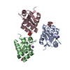
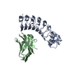
| Deposited unit | 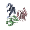
| ||||||||
|---|---|---|---|---|---|---|---|---|---|
| 1 |
| ||||||||
| Unit cell |
|
-Components
| #1: Protein | Design Mass: 14442.325 Da / Num. of mol.: 3 Source method: isolated from a genetically manipulated source Source: (gene. exp.) Trypanosoma brucei (eukaryote) / Production host:  Enterobacteria phage L1 (virus) Enterobacteria phage L1 (virus)#2: Water | ChemComp-HOH / | Water Mass: 18.015 Da / Num. of mol.: 83 / Source method: isolated from a natural source / Formula: H2O Mass: 18.015 Da / Num. of mol.: 83 / Source method: isolated from a natural source / Formula: H2O |
|---|
-Experimental details
-Experiment
| Experiment | Method: X-RAY DIFFRACTION / Number of used crystals: 1 |
|---|
- Sample preparation
| Crystal | Density Matthews: 2.63 Å3/Da / Density % sol: 53.21 % |
|---|---|
| Crystal grow | Temperature: 289 K / Method: vapor diffusion, sitting drop / pH: 5.6 Details: 0.2M Potassium sodium tartrate, 0.1M Sodium citrate pH5.6, 2.0MAmmonium sulfate |
-Data collection
| Diffraction | Mean temperature: 100 K / Serial crystal experiment: N |
|---|---|
| Diffraction source | Source: SYNCHROTRON / Site: SSRF / Beamline: BL17U1 / Wavelength: 0.9791 Å |
| Detector | Type: ADSC QUANTUM 315r / Detector: CCD / Date: May 21, 2017 |
| Radiation | Protocol: SINGLE WAVELENGTH / Monochromatic (M) / Laue (L): M / Scattering type: x-ray |
| Radiation wavelength | Wavelength: 0.9791 Å / Relative weight: 1 |
| Reflection | Resolution: 2.25→124.86 Å / Num. obs: 22476 / % possible obs: 99.8 % / Redundancy: 27.5 % / CC1/2: 0.999 / Rmerge(I) obs: 0.118 / Rpim(I) all: 0.023 / Rrim(I) all: 0.12 / Net I/av σ(I): 21.8 / Net I/σ(I): 21.8 |
| Reflection shell | Resolution: 2.25→2.31 Å / Redundancy: 28.9 % / Rmerge(I) obs: 0.663 / Mean I/σ(I) obs: 6.9 / Num. unique obs: 1628 / CC1/2: 0.977 / Rpim(I) all: 0.125 / Rrim(I) all: 0.675 / % possible all: 100 |
- Processing
| Software |
| |||||||||||||||||||||||||||||||||||||||||||||||||||||||||||||||
|---|---|---|---|---|---|---|---|---|---|---|---|---|---|---|---|---|---|---|---|---|---|---|---|---|---|---|---|---|---|---|---|---|---|---|---|---|---|---|---|---|---|---|---|---|---|---|---|---|---|---|---|---|---|---|---|---|---|---|---|---|---|---|---|---|
| Refinement | Method to determine structure: MOLECULAR REPLACEMENT Starting model: 1R26 Resolution: 2.25→44.143 Å / SU ML: 0.28 / Cross valid method: NONE / σ(F): 1.39 / Phase error: 25.28
| |||||||||||||||||||||||||||||||||||||||||||||||||||||||||||||||
| Solvent computation | Shrinkage radii: 0.9 Å / VDW probe radii: 1.11 Å | |||||||||||||||||||||||||||||||||||||||||||||||||||||||||||||||
| Refinement step | Cycle: LAST / Resolution: 2.25→44.143 Å
| |||||||||||||||||||||||||||||||||||||||||||||||||||||||||||||||
| Refine LS restraints |
| |||||||||||||||||||||||||||||||||||||||||||||||||||||||||||||||
| LS refinement shell |
| |||||||||||||||||||||||||||||||||||||||||||||||||||||||||||||||
| Refinement TLS params. | Method: refined / Origin x: 31.4312 Å / Origin y: 6.2188 Å / Origin z: 5.6131 Å
| |||||||||||||||||||||||||||||||||||||||||||||||||||||||||||||||
| Refinement TLS group | Selection details: all |
