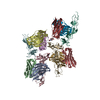
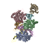
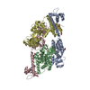
Crystal structure of DNA-free E.coli MutS P839E dimer mutant
Components
DNA mismatch repair protein MutS
Keywords
DNA BINDING PROTEIN / MutS / DNA mismatch repair / helix folding / ABC-ATPase / coiled coil / helix kink / apo-MutS
Function / homology
Function and homology information
adenine/cytosine mispair binding / MutS complex / mismatch repair complex / regulation of DNA recombination / mismatched DNA binding / DNA binding, bending / ATP-dependent DNA damage sensor activity / mismatch repair / ADP binding / damaged DNA binding ...adenine/cytosine mispair binding / MutS complex / mismatch repair complex / regulation of DNA recombination / mismatched DNA binding / DNA binding, bending / ATP-dependent DNA damage sensor activity / mismatch repair / ADP binding / damaged DNA binding / DNA damage response / ATP hydrolysis activity / ATP binding / identical protein binding / cytosol Similarity search - Function
MutS, connector domain / DNA repair protein MutS, domain I / MutS, DNA mismatch repair protein; Chain A, domain 3 / MutS, DNA mismatch repair protein; Chain A, domain 3 - #10 / DNA mismatch repair protein MutS / DNA mismatch repair protein MutS/MSH / DNA mismatch repair protein MutS-like, N-terminal / DNA mismatch repair protein MutS, connector domain / DNA mismatch repair protein MutS, clamp / DNA mismatch repair protein MutS, N-terminal ...MutS, connector domain / DNA repair protein MutS, domain I / MutS, DNA mismatch repair protein; Chain A, domain 3 / MutS, DNA mismatch repair protein; Chain A, domain 3 - #10 / DNA mismatch repair protein MutS / DNA mismatch repair protein MutS/MSH / DNA mismatch repair protein MutS-like, N-terminal / DNA mismatch repair protein MutS, connector domain / DNA mismatch repair protein MutS, clamp / DNA mismatch repair protein MutS, N-terminal / MutS, connector domain superfamily / MutS domain I / MutS domain II / MutS family domain IV / MutS domain III / DNA mismatch repair MutS family / DNA mismatch repair protein MutS, C-terminal / DNA mismatch repair protein MutS, core / DNA mismatch repair protein MutS, core domain superfamily / MutS domain V / DNA mismatch repair proteins mutS family signature. / DNA-binding domain of DNA mismatch repair MUTS family / ATPase domain of DNA mismatch repair MUTS family / MutS, DNA mismatch repair protein, domain I / Nucleotidyltransferase; domain 5 / P-loop containing nucleotide triphosphate hydrolases / P-loop containing nucleoside triphosphate hydrolase / Rossmann fold / 2-Layer Sandwich / Orthogonal Bundle / 3-Layer(aba) Sandwich / Mainly Alpha / Alpha Beta Similarity search - Domain/homology
Resolution: 2.6→71.61 Å / Cor.coef. Fo:Fc: 0.902 / Cor.coef. Fo:Fc free: 0.855 / SU R Cruickshank DPI: 0.54 / Cross valid method: THROUGHOUT / σ(F): 0 / SU R Blow DPI: 0.52 / SU Rfree Blow DPI: 0.294 / SU Rfree Cruickshank DPI: 0.301 Details: Residues 12-80 (mismatch binding domain) in chain A and residues 442-503 (clamp domain) in chain B are are likely flexible They were placed in the structure using conserved fold of the ...Details: Residues 12-80 (mismatch binding domain) in chain A and residues 442-503 (clamp domain) in chain B are are likely flexible They were placed in the structure using conserved fold of the mismatch binding domain and clamp domain, respectively
In the structure databanks used in Yorodumi, some data are registered as the other names, "COVID-19 virus" and "2019-nCoV". Here are the details of the virus and the list of structure data.
Jan 31, 2019. EMDB accession codes are about to change! (news from PDBe EMDB page)
EMDB accession codes are about to change! (news from PDBe EMDB page)
The allocation of 4 digits for EMDB accession codes will soon come to an end. Whilst these codes will remain in use, new EMDB accession codes will include an additional digit and will expand incrementally as the available range of codes is exhausted. The current 4-digit format prefixed with “EMD-” (i.e. EMD-XXXX) will advance to a 5-digit format (i.e. EMD-XXXXX), and so on. It is currently estimated that the 4-digit codes will be depleted around Spring 2019, at which point the 5-digit format will come into force.
The EM Navigator/Yorodumi systems omit the EMD- prefix.
Related info.:Q: What is EMD? / ID/Accession-code notation in Yorodumi/EM Navigator
Yorodumi is a browser for structure data from EMDB, PDB, SASBDB, etc.
This page is also the successor to EM Navigator detail page, and also detail information page/front-end page for Omokage search.
The word "yorodu" (or yorozu) is an old Japanese word meaning "ten thousand". "mi" (miru) is to see.
Related info.:EMDB / PDB / SASBDB / Comparison of 3 databanks / Yorodumi Search / Aug 31, 2016. New EM Navigator & Yorodumi / Yorodumi Papers / Jmol/JSmol / Function and homology information / Changes in new EM Navigator and Yorodumi
 Movie
Movie Controller


 Open data
 Basic information
 Components
 Keywords
 Function and homology information
 Authors
 Netherlands, 4items
  Citation
 Structure visualization
 Downloads & links
 Other downloads
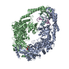


 PDBj
PDBj Assembly
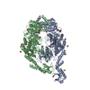

 Mass: 92.094 Da / Num. of mol.: 13 / Source method: obtained synthetically / Formula: C3H8O3
 Mass: 96.063 Da / Num. of mol.: 9 / Source method: obtained synthetically / Formula: SO4
 Mass: 427.201 Da / Num. of mol.: 2 / Source method: obtained synthetically / Formula: C10H15N5O10P2 / Comment: ADP, energy-carrying molecule*YM
 Mass: 18.015 Da / Num. of mol.: 140 / Source method: isolated from a natural source / Formula: H2O
 Sample preparation
 / Beamline: ID14-4 / Wavelength: 0.934 Å
 Processing