 Movie
Movie Controller

[English] 日本語
 Yorodumi
- PDB-6hzl: Crystal structure of redox-inhibited phosphoribulokinase from Syn... -
+ Open data

- Basic information
| Entry | Database: PDB / ID: 6hzl | ||||||
|---|---|---|---|---|---|---|---|
| Title | Crystal structure of redox-inhibited phosphoribulokinase from Synechococcus sp. (strain PCC 6301), osmate derivative | ||||||
 Components Components | Phosphoribulokinase | ||||||
 Keywords Keywords | TRANSFERASE / Phosphoribulokinase / Calvin cycle | ||||||
| Function / homology |  Function and homology informationphosphoribulokinase / phosphoribulokinase activity / carbohydrate metabolic process / phosphorylation / ATP binding Function and homology informationphosphoribulokinase / phosphoribulokinase activity / carbohydrate metabolic process / phosphorylation / ATP bindingSimilarity search - Function | ||||||
| Biological species |  Synechococcus elongatus PCC 6301 (bacteria) Synechococcus elongatus PCC 6301 (bacteria) | ||||||
| Method | X-RAY DIFFRACTION / SYNCHROTRON / SAD / Resolution: 2.77 Å | ||||||
 Authors Authors | Wilson, R.H. / Bracher, A. / Hartl, F.U. / Hayer-Hartl, M. | ||||||
 Citation Citation | Journal: Acta Crystallogr.,Sect.F / Year: 2019 Title: Crystal structure of phosphoribulokinase from Synechococcus sp. strain PCC 6301. Authors: Wilson, R.H. / Hayer-Hartl, M. / Bracher, A. | ||||||
| History |
|
- Structure visualization
| Structure viewer | Molecule: MolmilJmol/JSmol |
|---|
- Downloads & links
-Download
| PDBx/mmCIF format | 6hzl.cif.gz | 258.1 KB | Display | PDBx/mmCIF format |
|---|---|---|---|---|
| PDB format | pdb6hzl.ent.gz | 218.2 KB | Display | PDB format |
| PDBx/mmJSON format | 6hzl.json.gz | Tree view | PDBx/mmJSON format | |
| Others |  Other downloads Other downloads |
-Validation report
| Arichive directory | https://data.pdbj.org/pub/pdb/validation_reports/hz/6hzlftp://data.pdbj.org/pub/pdb/validation_reports/hz/6hzl | HTTPS FTP |
|---|
-Related structure data


-Links
 PDBj
PDBj- Assembly
| Deposited unit | 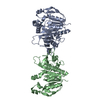
| |||||||||||||||||||||||||||
|---|---|---|---|---|---|---|---|---|---|---|---|---|---|---|---|---|---|---|---|---|---|---|---|---|---|---|---|---|
| 1 |
| |||||||||||||||||||||||||||
| Unit cell |
| |||||||||||||||||||||||||||
| Noncrystallographic symmetry (NCS) | NCS domain:
NCS domain segments:
|
-Components
| #1: Protein | Mass: 37760.750 Da / Num. of mol.: 2 Source method: isolated from a genetically manipulated source Source: (gene. exp.) Synechococcus elongatus PCC 6301 (bacteria)Gene: prk, syc0567_d / Plasmid: pHUE-prk4 / Production host: Escherichia coli BL21(DE3) (bacteria) / References: UniProt: A0A0H3K6J7, phosphoribulokinase#2: Chemical | Osmium  Mass: 190.230 Da / Num. of mol.: 2 / Source method: obtained synthetically / Formula: Os Mass: 190.230 Da / Num. of mol.: 2 / Source method: obtained synthetically / Formula: Os#3: Water | ChemComp-HOH / | Water Mass: 18.015 Da / Num. of mol.: 1 / Source method: isolated from a natural source / Formula: H2O Mass: 18.015 Da / Num. of mol.: 1 / Source method: isolated from a natural source / Formula: H2O |
|---|
-Experimental details
-Experiment
| Experiment | Method: X-RAY DIFFRACTION / Number of used crystals: 1 |
|---|
- Sample preparation
| Crystal | Density Matthews: 2.63 Å3/Da / Density % sol: 53.27 % |
|---|---|
| Crystal grow | Temperature: 277 K / Method: vapor diffusion / pH: 8 / Details: 20 % PEG-3350, 200 mM ammonium formate |
-Data collection
| Diffraction | Mean temperature: 100 K / Serial crystal experiment: N | ||||||||||||||||||||||||||||||
|---|---|---|---|---|---|---|---|---|---|---|---|---|---|---|---|---|---|---|---|---|---|---|---|---|---|---|---|---|---|---|---|
| Diffraction source | Source: SYNCHROTRON / Site: ESRF  / Beamline: ID29 / Wavelength: 1.13937 Å / Beamline: ID29 / Wavelength: 1.13937 Å | ||||||||||||||||||||||||||||||
| Detector | Type: DECTRIS PILATUS 6M / Detector: PIXEL / Date: Feb 4, 2017 | ||||||||||||||||||||||||||||||
| Radiation | Protocol: SINGLE WAVELENGTH / Monochromatic (M) / Laue (L): M / Scattering type: x-ray | ||||||||||||||||||||||||||||||
| Radiation wavelength | Wavelength: 1.13937 Å / Relative weight: 1 | ||||||||||||||||||||||||||||||
| Reflection | Resolution: 2.77→49.33 Å / Num. obs: 20468 / % possible obs: 100 % / Redundancy: 51.9 % / Biso Wilson estimate: 75 Å2 / CC1/2: 0.999 / Rmerge(I) obs: 0.226 / Rpim(I) all: 0.03 / Rrim(I) all: 0.228 / Net I/σ(I): 17.4 / Num. measured all: 1062487 / Scaling rejects: 15 | ||||||||||||||||||||||||||||||
| Reflection shell | Diffraction-ID: 1
|
-Phasing
| Phasing | Method: SAD |
|---|
- Processing
| Software |
| |||||||||||||||||||||||||||||||||||||||||||||||||||||||||||||||||||||||||||
|---|---|---|---|---|---|---|---|---|---|---|---|---|---|---|---|---|---|---|---|---|---|---|---|---|---|---|---|---|---|---|---|---|---|---|---|---|---|---|---|---|---|---|---|---|---|---|---|---|---|---|---|---|---|---|---|---|---|---|---|---|---|---|---|---|---|---|---|---|---|---|---|---|---|---|---|---|
| Refinement | Method to determine structure: SAD / Resolution: 2.77→30 Å / Cor.coef. Fo:Fc: 0.941 / Cor.coef. Fo:Fc free: 0.927 / WRfactor Rfree: 0.2532 / WRfactor Rwork: 0.2279 / FOM work R set: 0.7785 / SU B: 38.101 / SU ML: 0.33 / SU R Cruickshank DPI: 0.3879 / SU Rfree: 0.3686 / Cross valid method: THROUGHOUT / σ(F): 0 / ESU R Free: 0.369 / Stereochemistry target values: MAXIMUM LIKELIHOOD Details: HYDROGENS HAVE BEEN ADDED IN THE RIDING POSITIONS U VALUES : WITH TLS ADDED
| |||||||||||||||||||||||||||||||||||||||||||||||||||||||||||||||||||||||||||
| Solvent computation | Ion probe radii: 0.8 Å / Shrinkage radii: 0.8 Å / VDW probe radii: 1.2 Å / Solvent model: MASK | |||||||||||||||||||||||||||||||||||||||||||||||||||||||||||||||||||||||||||
| Displacement parameters | Biso max: 281.89 Å2 / Biso mean: 114.536 Å2 / Biso min: 52.6 Å2
| |||||||||||||||||||||||||||||||||||||||||||||||||||||||||||||||||||||||||||
| Refinement step | Cycle: final / Resolution: 2.77→30 Å
| |||||||||||||||||||||||||||||||||||||||||||||||||||||||||||||||||||||||||||
| Refine LS restraints |
| |||||||||||||||||||||||||||||||||||||||||||||||||||||||||||||||||||||||||||
| Refine LS restraints NCS | Ens-ID: 1 / Number: 18265 / Refine-ID: X-RAY DIFFRACTION / Type: interatomic distance / Rms dev position: 0.03 Å / Weight position: 0.05
| |||||||||||||||||||||||||||||||||||||||||||||||||||||||||||||||||||||||||||
| LS refinement shell | Resolution: 2.77→2.842 Å / Rfactor Rfree error: 0 / Total num. of bins used: 20
| |||||||||||||||||||||||||||||||||||||||||||||||||||||||||||||||||||||||||||
| Refinement TLS params. | Method: refined / Refine-ID: X-RAY DIFFRACTION
| |||||||||||||||||||||||||||||||||||||||||||||||||||||||||||||||||||||||||||
| Refinement TLS group |
|
