 Movie
Movie Controller

[English] 日本語
 Yorodumi
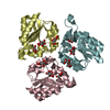
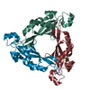
- PDB-6hsa: The crystal structure of type II Dehydroquinase from Butyrivibrio... -
+ Open data

- Basic information
| Entry | Database: PDB / ID: 6hsa | |||||||||
|---|---|---|---|---|---|---|---|---|---|---|
| Title | The crystal structure of type II Dehydroquinase from Butyrivibrio crossotus DSM 2876 | |||||||||
 Components Components | 3-dehydroquinate dehydratase | |||||||||
 Keywords Keywords | BIOSYNTHETIC PROTEIN / shikimate pathway / dehydratase | |||||||||
| Function / homology |  Function and homology information3-dehydroquinate dehydratase / 3-dehydroquinate dehydratase activity / chorismate biosynthetic process / aromatic amino acid family biosynthetic process / amino acid biosynthetic process Function and homology information3-dehydroquinate dehydratase / 3-dehydroquinate dehydratase activity / chorismate biosynthetic process / aromatic amino acid family biosynthetic process / amino acid biosynthetic processSimilarity search - Function | |||||||||
| Biological species |  Butyrivibrio crossotus DSM 2876 (bacteria) Butyrivibrio crossotus DSM 2876 (bacteria) | |||||||||
| Method | X-RAY DIFFRACTION / SYNCHROTRON / MOLECULAR REPLACEMENT / Resolution: 0.92 Å | |||||||||
 Authors Authors | Lapthorn, A.J. / Roszak, A.W. | |||||||||
| Funding support |  United Kingdom, 1items United Kingdom, 1items
| |||||||||
 Citation Citation | Journal: To Be Published Title: The crystal structure of type II Dehydroquinase from Butyrivibrio crossotus DSM 2876 Authors: Lapthorn, A.J. / Ner, L. / Roszak, A.W. #1: Journal: AMB Express / Year: 2015 Title: Unraveling the kinetic diversity of microbial 3-dehydroquinate dehydratases of shikimate pathway. Authors: Liu, C. / Liu, Y.M. / Sun, Q.L. / Jiang, C.Y. / Liu, S.J. #2: Journal: Structure / Year: 2002Title: The structure and mechanism of the type II dehydroquinase from Streptomyces coelicolor. Authors: Roszak, A.W. / Robinson, D.A. / Krell, T. / Hunter, I.S. / Fredrickson, M. / Abell, C. / Coggins, J.R. / Lapthorn, A.J. | |||||||||
| History |
|
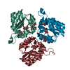
- Structure visualization
| Structure viewer | Molecule: MolmilJmol/JSmol |
|---|
- Downloads & links
-Download
| PDBx/mmCIF format | 6hsa.cif.gz | 146.6 KB | Display | PDBx/mmCIF format |
|---|---|---|---|---|
| PDB format | pdb6hsa.ent.gz | 116.1 KB | Display | PDB format |
| PDBx/mmJSON format | 6hsa.json.gz | Tree view | PDBx/mmJSON format | |
| Others |  Other downloads Other downloads |
-Validation report
| Arichive directory | https://data.pdbj.org/pub/pdb/validation_reports/hs/6hsaftp://data.pdbj.org/pub/pdb/validation_reports/hs/6hsa | HTTPS FTP |
|---|
-Related structure data
| Related structure data | 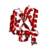 6hs8C 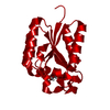 6hs9SC 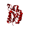 6hsbC  6hsqC S: Starting model for refinement C: citing same article ( |
|---|---|
| Similar structure data |


-Links
 PDBj
PDBj


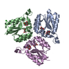
- Assembly
| Deposited unit | 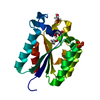
| |||||||||||||||||||||||||||
|---|---|---|---|---|---|---|---|---|---|---|---|---|---|---|---|---|---|---|---|---|---|---|---|---|---|---|---|---|
| 1 | 
| |||||||||||||||||||||||||||
| Unit cell |
| |||||||||||||||||||||||||||
| Components on special symmetry positions |
|
-Components
-Protein , 1 types, 1 molecules A
| #1: Protein | / 3-dehydroquinase / Type II DHQase Mass: 16387.510 Da / Num. of mol.: 1 Source method: isolated from a genetically manipulated source Source: (gene. exp.) Butyrivibrio crossotus DSM 2876 (bacteria)Gene: aroQ, BUTYVIB_01550 / Plasmid: pET28a+ / Production host: Escherichia coli BL21(DE3) (bacteria) / References: UniProt: D4S0D1, 3-dehydroquinate dehydratase |
|---|
-Non-polymers , 6 types, 283 molecules 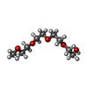


| #2: Chemical | ChemComp-ETE /  Mass: 208.252 Da / Num. of mol.: 1 / Source method: obtained synthetically / Formula: C9H20O5 Mass: 208.252 Da / Num. of mol.: 1 / Source method: obtained synthetically / Formula: C9H20O5 | ||||||
|---|---|---|---|---|---|---|---|
| #3: Chemical | ChemComp-SER / Serine Type: L-peptide linking / Mass: 105.093 Da / Num. of mol.: 1 / Source method: obtained synthetically / Formula: C3H7NO3 Type: L-peptide linking / Mass: 105.093 Da / Num. of mol.: 1 / Source method: obtained synthetically / Formula: C3H7NO3 | ||||||
| #4: Chemical | Sulfate Mass: 96.063 Da / Num. of mol.: 2 / Source method: obtained synthetically / Formula: SO4 Mass: 96.063 Da / Num. of mol.: 2 / Source method: obtained synthetically / Formula: SO4#5: Chemical | ChemComp-FLC / | Citric acid Mass: 189.100 Da / Num. of mol.: 1 / Source method: obtained synthetically / Formula: C6H5O7 Mass: 189.100 Da / Num. of mol.: 1 / Source method: obtained synthetically / Formula: C6H5O7#6: Chemical | ChemComp-GOL / | Glycerol Mass: 92.094 Da / Num. of mol.: 1 / Source method: obtained synthetically / Formula: C3H8O3 Mass: 92.094 Da / Num. of mol.: 1 / Source method: obtained synthetically / Formula: C3H8O3#7: Water | ChemComp-HOH / | WaterMass: 18.015 Da / Num. of mol.: 277 / Source method: isolated from a natural source / Formula: H2O |
-Experimental details
-Experiment
| Experiment | Method: X-RAY DIFFRACTION / Number of used crystals: 1 |
|---|
- Sample preparation
| Crystal | Density Matthews: 2.67 Å3/Da / Density % sol: 53.96 % / Description: hexagonal prism |
|---|---|
| Crystal grow | Temperature: 293 K / Method: vapor diffusion, sitting drop / pH: 5.6 Details: 20% PEG 4000, 0.2M Ammonium sulfate, 0.1M sodium citrate pH 5.6 |
-Data collection
| Diffraction | Mean temperature: 100 K | |||||||||||||||||||||||||||
|---|---|---|---|---|---|---|---|---|---|---|---|---|---|---|---|---|---|---|---|---|---|---|---|---|---|---|---|---|
| Diffraction source | Source: SYNCHROTRON / Site: Diamond / Beamline: I03 / Wavelength: 0.82656 Å | |||||||||||||||||||||||||||
| Detector | Type: DECTRIS PILATUS3 S 6M / Detector: PIXEL / Date: Dec 18, 2016 | |||||||||||||||||||||||||||
| Radiation | Protocol: SINGLE WAVELENGTH / Monochromatic (M) / Laue (L): M / Scattering type: x-ray | |||||||||||||||||||||||||||
| Radiation wavelength | Wavelength: 0.82656 Å / Relative weight: 1 | |||||||||||||||||||||||||||
| Reflection | Resolution: 0.92→31.93 Å / Num. obs: 112975 / % possible obs: 96.7 % / Redundancy: 3.2 % / Biso Wilson estimate: 6.6 Å2 / CC1/2: 0.997 / Rmerge(I) obs: 0.046 / Rpim(I) all: 0.03 / Rsym value: 0.055 / Net I/σ(I): 14.1 | |||||||||||||||||||||||||||
| Reflection shell | Diffraction-ID: 1
|
- Processing
| Software |
| |||||||||||||||||||||||||||||||||||||||||||||||||||||||||||||||||||||||||||
|---|---|---|---|---|---|---|---|---|---|---|---|---|---|---|---|---|---|---|---|---|---|---|---|---|---|---|---|---|---|---|---|---|---|---|---|---|---|---|---|---|---|---|---|---|---|---|---|---|---|---|---|---|---|---|---|---|---|---|---|---|---|---|---|---|---|---|---|---|---|---|---|---|---|---|---|---|
| Refinement | Method to determine structure: MOLECULAR REPLACEMENT Starting model: 6HS9 Resolution: 0.92→31.93 Å / Cor.coef. Fo:Fc: 0.987 / Cor.coef. Fo:Fc free: 0.981 / SU B: 0.29 / SU ML: 0.008 / Cross valid method: THROUGHOUT / σ(F): 0 / ESU R: 0.012 / ESU R Free: 0.014 Details: U VALUES : WITH TLS ADDED HYDROGENS HAVE BEEN USED IF PRESENT IN THE INPUT
| |||||||||||||||||||||||||||||||||||||||||||||||||||||||||||||||||||||||||||
| Solvent computation | Ion probe radii: 0.8 Å / Shrinkage radii: 0.8 Å / VDW probe radii: 1.2 Å | |||||||||||||||||||||||||||||||||||||||||||||||||||||||||||||||||||||||||||
| Displacement parameters | Biso max: 89.07 Å2 / Biso mean: 12.663 Å2 / Biso min: 4.55 Å2
| |||||||||||||||||||||||||||||||||||||||||||||||||||||||||||||||||||||||||||
| Refinement step | Cycle: final / Resolution: 0.92→31.93 Å
| |||||||||||||||||||||||||||||||||||||||||||||||||||||||||||||||||||||||||||
| Refine LS restraints |
| |||||||||||||||||||||||||||||||||||||||||||||||||||||||||||||||||||||||||||
| LS refinement shell | Resolution: 0.923→0.947 Å / Rfactor Rfree error: 0 / Total num. of bins used: 20
| |||||||||||||||||||||||||||||||||||||||||||||||||||||||||||||||||||||||||||
| Refinement TLS params. | Method: refined / Origin x: 26.078 Å / Origin y: -12.113 Å / Origin z: 12.96 Å
|
