 Movie
Movie Controller


+ Open data

- Basic information
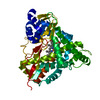
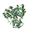
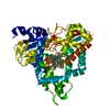
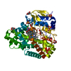
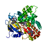
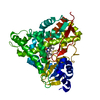
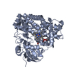
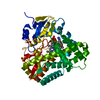
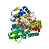
| Entry | Database: PDB / ID: 6hqm | ||||||||||||
|---|---|---|---|---|---|---|---|---|---|---|---|---|---|
| Title | Crystal structure of GcoA F169I bound to guaiacol | ||||||||||||
 Components Components | Cytochrome P450 | ||||||||||||
 Keywords Keywords | OXIDOREDUCTASE / cytochrome / P450 / lignin. | ||||||||||||
| Function / homology |  Function and homology informationOxidoreductases; Acting on paired donors, with incorporation or reduction of molecular oxygen; With reduced flavin or flavoprotein as one donor, and incorporation of one atom of oxygen into the other donor / : / oxidoreductase activity, acting on paired donors, with incorporation or reduction of molecular oxygen / monooxygenase activity / iron ion binding / heme binding Function and homology informationOxidoreductases; Acting on paired donors, with incorporation or reduction of molecular oxygen; With reduced flavin or flavoprotein as one donor, and incorporation of one atom of oxygen into the other donor / : / oxidoreductase activity, acting on paired donors, with incorporation or reduction of molecular oxygen / monooxygenase activity / iron ion binding / heme bindingSimilarity search - Function | ||||||||||||
| Biological species |  Amycolatopsis sp. ATCC 39116 (bacteria) Amycolatopsis sp. ATCC 39116 (bacteria) | ||||||||||||
| Method | X-RAY DIFFRACTION / SYNCHROTRON / MOLECULAR REPLACEMENT / Resolution: 1.85 Å | ||||||||||||
 Authors Authors | Mallinson, S.J.B. / Hinchen, D.J. / Allen, M.D. / Johnson, C.W. / Beckham, G.T. / McGeehan, J.E. | ||||||||||||
| Funding support |  United Kingdom, United Kingdom,  United States, 3items United States, 3items
| ||||||||||||
 Citation Citation | Journal: Proc.Natl.Acad.Sci.USA / Year: 2019 Title: Enabling microbial syringol conversion through structure-guided protein engineering. Authors: Machovina, M.M. / Mallinson, S.J.B. / Knott, B.C. / Meyers, A.W. / Garcia-Borras, M. / Bu, L. / Gado, J.E. / Oliver, A. / Schmidt, G.P. / Hinchen, D.J. / Crowley, M.F. / Johnson, C.W. / ...Authors: Machovina, M.M. / Mallinson, S.J.B. / Knott, B.C. / Meyers, A.W. / Garcia-Borras, M. / Bu, L. / Gado, J.E. / Oliver, A. / Schmidt, G.P. / Hinchen, D.J. / Crowley, M.F. / Johnson, C.W. / Neidle, E.L. / Payne, C.M. / Houk, K.N. / Beckham, G.T. / McGeehan, J.E. / DuBois, J.L. | ||||||||||||
| History |
|
- Structure visualization
| Structure viewer | Molecule: MolmilJmol/JSmol |
|---|
- Downloads & links
-Download
| PDBx/mmCIF format | 6hqm.cif.gz | 190.9 KB | Display | PDBx/mmCIF format |
|---|---|---|---|---|
| PDB format | pdb6hqm.ent.gz | 149.7 KB | Display | PDB format |
| PDBx/mmJSON format | 6hqm.json.gz | Tree view | PDBx/mmJSON format | |
| Others |  Other downloads Other downloads |
-Validation report
| Arichive directory | https://data.pdbj.org/pub/pdb/validation_reports/hq/6hqmftp://data.pdbj.org/pub/pdb/validation_reports/hq/6hqm | HTTPS FTP |
|---|
-Related structure data
| Related structure data |  6hqkC 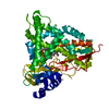 6hqlC 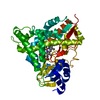 6hqnC 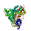 6hqoC 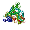 6hqpC  6hqqC  6hqrC 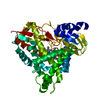 6hqsC 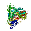 6hqtC  5ncbS S: Starting model for refinement C: citing same article ( |
|---|---|
| Similar structure data |

-Links
 PDBj
PDBj
- Assembly
| Deposited unit | 
| |||||||||
|---|---|---|---|---|---|---|---|---|---|---|
| 1 |
| |||||||||
| Unit cell |
| |||||||||
| Components on special symmetry positions |
|
-Components
| #1: Protein | Mass: 45418.762 Da / Num. of mol.: 1 / Mutation: F169I Source method: isolated from a genetically manipulated source Source: (gene. exp.) Amycolatopsis sp. ATCC 39116 (bacteria)Gene: AMETH_3834 / Production host: Escherichia coli (E. coli) / References: UniProt: A0A076MY51, UniProt: P0DPQ7*PLUS |
|---|---|
| #2: Chemical | ChemComp-HEM / Heme B  Mass: 616.487 Da / Num. of mol.: 1 / Source method: obtained synthetically / Formula: C34H32FeN4O4 Mass: 616.487 Da / Num. of mol.: 1 / Source method: obtained synthetically / Formula: C34H32FeN4O4 |
| #3: Chemical | ChemComp-JZ3 / Guaiacol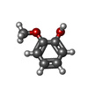  Mass: 124.137 Da / Num. of mol.: 1 / Source method: obtained synthetically / Formula: C7H8O2 / Feature type: SUBJECT OF INVESTIGATION Mass: 124.137 Da / Num. of mol.: 1 / Source method: obtained synthetically / Formula: C7H8O2 / Feature type: SUBJECT OF INVESTIGATION |
| #4: Water | ChemComp-HOH / Water Mass: 18.015 Da / Num. of mol.: 426 / Source method: isolated from a natural source / Formula: H2O Mass: 18.015 Da / Num. of mol.: 426 / Source method: isolated from a natural source / Formula: H2O |
-Experimental details
-Experiment
| Experiment | Method: X-RAY DIFFRACTION / Number of used crystals: 1 |
|---|
- Sample preparation
| Crystal | Density Matthews: 3.43 Å3/Da / Density % sol: 64.13 % |
|---|---|
| Crystal grow | Temperature: 289 K / Method: vapor diffusion, hanging drop / Details: Sodium malonate, HEPES, guaiacol. |
-Data collection
| Diffraction | Mean temperature: 100 K |
|---|---|
| Diffraction source | Source: SYNCHROTRON / Site: Diamond / Beamline: I04 / Wavelength: 0.9795 Å |
| Detector | Type: DECTRIS PILATUS 6M-F / Detector: PIXEL / Date: May 20, 2018 |
| Radiation | Protocol: SINGLE WAVELENGTH / Monochromatic (M) / Laue (L): M / Scattering type: x-ray |
| Radiation wavelength | Wavelength: 0.9795 Å / Relative weight: 1 |
| Reflection | Resolution: 1.85→56.25 Å / Num. obs: 54123 / % possible obs: 100 % / Redundancy: 12.7 % / CC1/2: 1 / Net I/σ(I): 10.4 |
| Reflection shell | Resolution: 1.85→1.9 Å / Mean I/σ(I) obs: 2.6 / CC1/2: 0.849 |
- Processing
| Software |
| ||||||||||||||||||||||||||||||||||||||||||||||||||||||||||||||||||||||||||||||||||||||||||||||||||||||||||||||||||||||||||||||||||||||||||||
|---|---|---|---|---|---|---|---|---|---|---|---|---|---|---|---|---|---|---|---|---|---|---|---|---|---|---|---|---|---|---|---|---|---|---|---|---|---|---|---|---|---|---|---|---|---|---|---|---|---|---|---|---|---|---|---|---|---|---|---|---|---|---|---|---|---|---|---|---|---|---|---|---|---|---|---|---|---|---|---|---|---|---|---|---|---|---|---|---|---|---|---|---|---|---|---|---|---|---|---|---|---|---|---|---|---|---|---|---|---|---|---|---|---|---|---|---|---|---|---|---|---|---|---|---|---|---|---|---|---|---|---|---|---|---|---|---|---|---|---|---|---|
| Refinement | Method to determine structure: MOLECULAR REPLACEMENT Starting model: 5NCB Resolution: 1.85→52.605 Å / SU ML: 0.21 / Cross valid method: FREE R-VALUE / σ(F): 1.33 / Phase error: 19.6
| ||||||||||||||||||||||||||||||||||||||||||||||||||||||||||||||||||||||||||||||||||||||||||||||||||||||||||||||||||||||||||||||||||||||||||||
| Solvent computation | Shrinkage radii: 0.9 Å / VDW probe radii: 1.11 Å | ||||||||||||||||||||||||||||||||||||||||||||||||||||||||||||||||||||||||||||||||||||||||||||||||||||||||||||||||||||||||||||||||||||||||||||
| Refinement step | Cycle: LAST / Resolution: 1.85→52.605 Å
| ||||||||||||||||||||||||||||||||||||||||||||||||||||||||||||||||||||||||||||||||||||||||||||||||||||||||||||||||||||||||||||||||||||||||||||
| Refine LS restraints |
| ||||||||||||||||||||||||||||||||||||||||||||||||||||||||||||||||||||||||||||||||||||||||||||||||||||||||||||||||||||||||||||||||||||||||||||
| LS refinement shell |
| ||||||||||||||||||||||||||||||||||||||||||||||||||||||||||||||||||||||||||||||||||||||||||||||||||||||||||||||||||||||||||||||||||||||||||||
| Refinement TLS params. | Method: refined / Origin x: 46.9146 Å / Origin y: 81.6712 Å / Origin z: 41.3709 Å
| ||||||||||||||||||||||||||||||||||||||||||||||||||||||||||||||||||||||||||||||||||||||||||||||||||||||||||||||||||||||||||||||||||||||||||||
| Refinement TLS group | Selection details: all |
