 Movie
Movie Controller

[English] 日本語
 Yorodumi
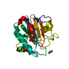
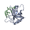
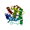
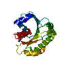
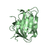
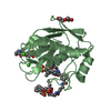
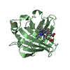
- PDB-6hkq: Human GPX4 in complex with covalent Inhibitor ML162 (S enantiomer) -
+ Open data

- Basic information
| Entry | Database: PDB / ID: 6hkq | ||||||
|---|---|---|---|---|---|---|---|
| Title | Human GPX4 in complex with covalent Inhibitor ML162 (S enantiomer) | ||||||
 Components Components | Phospholipid hydroperoxide glutathione peroxidase | ||||||
 Keywords Keywords |  OXIDOREDUCTASE / ANTI-OXIDATVE DEFENSE SYSTEM / Covalent inhibitor OXIDOREDUCTASE / ANTI-OXIDATVE DEFENSE SYSTEM / Covalent inhibitor | ||||||
| Function / homology |  Function and homology informationphospholipid-hydroperoxide glutathione peroxidase / phospholipid-hydroperoxide glutathione peroxidase activity / Synthesis of 15-eicosatetraenoic acid derivatives / Synthesis of 12-eicosatetraenoic acid derivatives / negative regulation of ferroptosis / selenium binding / glutathione peroxidase / Synthesis of 5-eicosatetraenoic acids / lipoxygenase pathway / arachidonic acid metabolic process ...phospholipid-hydroperoxide glutathione peroxidase / phospholipid-hydroperoxide glutathione peroxidase activity / Synthesis of 15-eicosatetraenoic acid derivatives / Synthesis of 12-eicosatetraenoic acid derivatives / negative regulation of ferroptosis / selenium binding / glutathione peroxidase / Synthesis of 5-eicosatetraenoic acids / lipoxygenase pathway / arachidonic acid metabolic process / Biosynthesis of aspirin-triggered D-series resolvins / Biosynthesis of E-series 18(R)-resolvins / Biosynthesis of D-series resolvins / Biosynthesis of E-series 18(S)-resolvins / glutathione peroxidase activity / long-chain fatty acid biosynthetic process / protein polymerization / phospholipid metabolic process / response to estradiol / nuclear envelope / chromatin organization / cellular response to oxidative stress / spermatogenesis / response to oxidative stress / protein-containing complex / mitochondrion / extracellular exosome / identical protein binding / nucleus / cytosol Function and homology informationphospholipid-hydroperoxide glutathione peroxidase / phospholipid-hydroperoxide glutathione peroxidase activity / Synthesis of 15-eicosatetraenoic acid derivatives / Synthesis of 12-eicosatetraenoic acid derivatives / negative regulation of ferroptosis / selenium binding / glutathione peroxidase / Synthesis of 5-eicosatetraenoic acids / lipoxygenase pathway / arachidonic acid metabolic process ...phospholipid-hydroperoxide glutathione peroxidase / phospholipid-hydroperoxide glutathione peroxidase activity / Synthesis of 15-eicosatetraenoic acid derivatives / Synthesis of 12-eicosatetraenoic acid derivatives / negative regulation of ferroptosis / selenium binding / glutathione peroxidase / Synthesis of 5-eicosatetraenoic acids / lipoxygenase pathway / arachidonic acid metabolic process / Biosynthesis of aspirin-triggered D-series resolvins / Biosynthesis of E-series 18(R)-resolvins / Biosynthesis of D-series resolvins / Biosynthesis of E-series 18(S)-resolvins / glutathione peroxidase activity / long-chain fatty acid biosynthetic process / protein polymerization / phospholipid metabolic process / response to estradiol / nuclear envelope / chromatin organization / cellular response to oxidative stress / spermatogenesis / response to oxidative stress / protein-containing complex / mitochondrion / extracellular exosome / identical protein binding / nucleus / cytosolSimilarity search - Function | ||||||
| Biological species |  Homo sapiens (human) Homo sapiens (human) | ||||||
| Method | X-RAY DIFFRACTION / MOLECULAR REPLACEMENT / Resolution: 1.54 Å | ||||||
 Authors Authors | Hillig, R.C. / Moosmayer, D. / Hilpmann, A. / Hoffmann, L. / Schnirch, L. / Eaton, J.K. / Badock, V. / Gradl, S. | ||||||
 Citation Citation | Journal: Acta Crystallogr D Struct Biol / Year: 2021 Title: Crystal structures of the selenoprotein glutathione peroxidase 4 in its apo form and in complex with the covalently bound inhibitor ML162. Authors: Moosmayer, D. / Hilpmann, A. / Hoffmann, J. / Schnirch, L. / Zimmermann, K. / Badock, V. / Furst, L. / Eaton, J.K. / Viswanathan, V.S. / Schreiber, S.L. / Gradl, S. / Hillig, R.C. | ||||||
| History |
|
- Structure visualization
| Structure viewer | Molecule: MolmilJmol/JSmol |
|---|
- Downloads & links
-Download
| PDBx/mmCIF format | 6hkq.cif.gz | 83.8 KB | Display | PDBx/mmCIF format |
|---|---|---|---|---|
| PDB format | pdb6hkq.ent.gz | 64.3 KB | Display | PDB format |
| PDBx/mmJSON format | 6hkq.json.gz | Tree view | PDBx/mmJSON format | |
| Others |  Other downloads Other downloads |
-Validation report
| Arichive directory | https://data.pdbj.org/pub/pdb/validation_reports/hk/6hkqftp://data.pdbj.org/pub/pdb/validation_reports/hk/6hkq | HTTPS FTP |
|---|
-Related structure data


-Links
 PDBj
PDBj
- Assembly
| Deposited unit | 
| ||||||||
|---|---|---|---|---|---|---|---|---|---|
| 1 |
| ||||||||
| Unit cell |
|
-Components
-Protein , 1 types, 1 molecules A
| #1: Protein | Mass: 20366.230 Da / Num. of mol.: 1 / Mutation: C66S Source method: isolated from a genetically manipulated source Details: C-terminal hexa-His tag added for affinity chromatography Source: (gene. exp.) Homo sapiens (human) / Gene: GPX4 / Production host:  Escherichia coli (E. coli) Escherichia coli (E. coli)References: UniProt: P36969, phospholipid-hydroperoxide glutathione peroxidase |
|---|
-Non-polymers , 5 types, 232 molecules 


| #2: Chemical | ChemComp-G9N / ( Mass: 477.403 Da / Num. of mol.: 1 / Source method: obtained synthetically / Formula: C23H22Cl2N2O3S / Feature type: SUBJECT OF INVESTIGATION Mass: 477.403 Da / Num. of mol.: 1 / Source method: obtained synthetically / Formula: C23H22Cl2N2O3S / Feature type: SUBJECT OF INVESTIGATION | ||
|---|---|---|---|
| #3: Chemical | ChemComp-SO4 / Sulfate Mass: 96.063 Da / Num. of mol.: 1 / Source method: obtained synthetically / Formula: SO4 Mass: 96.063 Da / Num. of mol.: 1 / Source method: obtained synthetically / Formula: SO4 | ||
| #4: Chemical | ChemComp-EDO / Ethylene glycol Mass: 62.068 Da / Num. of mol.: 1 / Source method: obtained synthetically / Formula: C2H6O2 Mass: 62.068 Da / Num. of mol.: 1 / Source method: obtained synthetically / Formula: C2H6O2 | ||
| #5: Chemical | Dimethyl sulfoxide Mass: 78.133 Da / Num. of mol.: 2 / Source method: obtained synthetically / Formula: C2H6OS / Comment: DMSO, precipitant*YM Mass: 78.133 Da / Num. of mol.: 2 / Source method: obtained synthetically / Formula: C2H6OS / Comment: DMSO, precipitant*YM#6: Water | ChemComp-HOH / | WaterMass: 18.015 Da / Num. of mol.: 227 / Source method: isolated from a natural source / Formula: H2O |
-Experimental details
-Experiment
| Experiment | Method: X-RAY DIFFRACTION / Number of used crystals: 1 |
|---|
- Sample preparation
| Crystal | Density Matthews: 1.87 Å3/Da / Density % sol: 34.24 % |
|---|---|
| Crystal grow | Temperature: 293 K / Method: vapor diffusion, hanging drop / pH: 8 Details: Reservoir made from 0.2 M ammonium sulfate, 20% (w/v) PEG 3350. Prio to crystal set-up, the protein was modified with covalent inhibitor, purified via gelfiltration and concentrated to 13.5 ...Details: Reservoir made from 0.2 M ammonium sulfate, 20% (w/v) PEG 3350. Prio to crystal set-up, the protein was modified with covalent inhibitor, purified via gelfiltration and concentrated to 13.5 mg/ml in 50 mM TrisHCl pH 8.0, 150 mM NaCl, 5mM TCEP |
-Data collection
| Diffraction | Mean temperature: 100 K |
|---|---|
| Diffraction source | Source: ROTATING ANODE / Type: RIGAKU MICROMAX-007 / Wavelength: 1.5418 Å |
| Detector | Type: DECTRIS PILATUS 200K / Detector: PIXEL / Date: Mar 26, 2018 |
| Radiation | Protocol: SINGLE WAVELENGTH / Monochromatic (M) / Laue (L): M / Scattering type: x-ray |
| Radiation wavelength | Wavelength: 1.5418 Å / Relative weight: 1 |
| Reflection | Resolution: 1.54→46.81 Å / Num. obs: 20009 / % possible obs: 86.6 % / Redundancy: 10.5 % / Rpim(I) all: 0.03 / Rrim(I) all: 0.11 / Rsym value: 0.1 / Net I/σ(I): 20.2 |
| Reflection shell | Resolution: 1.54→1.59 Å / Redundancy: 6.7 % / Mean I/σ(I) obs: 2.4 / Num. unique obs: 1092 / CC1/2: 0.81 / Rpim(I) all: 0.33 / Rrim(I) all: 0.97 / Rsym value: 0.9 / % possible all: 72.1 |
- Processing
| Software |
| ||||||||||||||||||||||||||||||||||||||||||||||||||||||||||||||||||||||||||||||||||||||||||||||||||||||||||||||||||||||||||||||||||||||||||||||||||||||||||||||||||||||||||||||||||||||
|---|---|---|---|---|---|---|---|---|---|---|---|---|---|---|---|---|---|---|---|---|---|---|---|---|---|---|---|---|---|---|---|---|---|---|---|---|---|---|---|---|---|---|---|---|---|---|---|---|---|---|---|---|---|---|---|---|---|---|---|---|---|---|---|---|---|---|---|---|---|---|---|---|---|---|---|---|---|---|---|---|---|---|---|---|---|---|---|---|---|---|---|---|---|---|---|---|---|---|---|---|---|---|---|---|---|---|---|---|---|---|---|---|---|---|---|---|---|---|---|---|---|---|---|---|---|---|---|---|---|---|---|---|---|---|---|---|---|---|---|---|---|---|---|---|---|---|---|---|---|---|---|---|---|---|---|---|---|---|---|---|---|---|---|---|---|---|---|---|---|---|---|---|---|---|---|---|---|---|---|---|---|---|---|
| Refinement | Method to determine structure: MOLECULAR REPLACEMENT / Resolution: 1.54→46.81 Å / Cor.coef. Fo:Fc: 0.969 / Cor.coef. Fo:Fc free: 0.957 / SU B: 2.902 / SU ML: 0.052 / Cross valid method: THROUGHOUT / ESU R: 0.09 / ESU R Free: 0.089 / Stereochemistry target values: MAXIMUM LIKELIHOOD / Details: HYDROGENS HAVE BEEN ADDED IN THE RIDING POSITIONS
| ||||||||||||||||||||||||||||||||||||||||||||||||||||||||||||||||||||||||||||||||||||||||||||||||||||||||||||||||||||||||||||||||||||||||||||||||||||||||||||||||||||||||||||||||||||||
| Solvent computation | Ion probe radii: 0.8 Å / Shrinkage radii: 0.8 Å / VDW probe radii: 1.2 Å / Solvent model: BABINET MODEL WITH MASK | ||||||||||||||||||||||||||||||||||||||||||||||||||||||||||||||||||||||||||||||||||||||||||||||||||||||||||||||||||||||||||||||||||||||||||||||||||||||||||||||||||||||||||||||||||||||
| Displacement parameters | Biso mean: 18.524 Å2
| ||||||||||||||||||||||||||||||||||||||||||||||||||||||||||||||||||||||||||||||||||||||||||||||||||||||||||||||||||||||||||||||||||||||||||||||||||||||||||||||||||||||||||||||||||||||
| Refinement step | Cycle: 1 / Resolution: 1.54→46.81 Å
| ||||||||||||||||||||||||||||||||||||||||||||||||||||||||||||||||||||||||||||||||||||||||||||||||||||||||||||||||||||||||||||||||||||||||||||||||||||||||||||||||||||||||||||||||||||||
| Refine LS restraints |
|