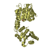
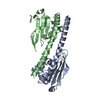
 Movie
Movie Controller


+ Open data

- Basic information
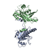
| Entry | Database: PDB / ID: 6hjo | ||||||
|---|---|---|---|---|---|---|---|
| Title | Myxococcus xanthus MglA bound to GDP | ||||||
 Components Components | Mutual gliding-motility protein MglA | ||||||
 Keywords Keywords |  CYTOSOLIC PROTEIN / Small GTPase CYTOSOLIC PROTEIN / Small GTPase | ||||||
| Function / homology |  Function and homology informationregulation of protein localization / GTPase activity / GTP binding / cytoplasm Function and homology informationregulation of protein localization / GTPase activity / GTP binding / cytoplasmSimilarity search - Function | ||||||
| Biological species |  Myxococcus xanthus DK 1622 (bacteria) Myxococcus xanthus DK 1622 (bacteria) | ||||||
| Method | X-RAY DIFFRACTION / SYNCHROTRON / MOLECULAR REPLACEMENT / Resolution: 1.98 Å | ||||||
 Authors Authors | Varela, P.F. / Cherfils, J. | ||||||
 Citation Citation | Journal: Nat Commun / Year: 2019 Title: MglA functions as a three-state GTPase to control movement reversals of Myxococcus xanthus. Authors: Galicia, C. / Lhospice, S. / Varela, P.F. / Trapani, S. / Zhang, W. / Navaza, J. / Herrou, J. / Mignot, T. / Cherfils, J. | ||||||
| History |
|
- Structure visualization
| Structure viewer | Molecule: MolmilJmol/JSmol |
|---|
- Downloads & links
-Download
| PDBx/mmCIF format | 6hjo.cif.gz | 169.5 KB | Display | PDBx/mmCIF format |
|---|---|---|---|---|
| PDB format | pdb6hjo.ent.gz | 134.6 KB | Display | PDB format |
| PDBx/mmJSON format | 6hjo.json.gz | Tree view | PDBx/mmJSON format | |
| Others |  Other downloads Other downloads |
-Validation report
| Arichive directory | https://data.pdbj.org/pub/pdb/validation_reports/hj/6hjoftp://data.pdbj.org/pub/pdb/validation_reports/hj/6hjo | HTTPS FTP |
|---|
-Related structure data
| Related structure data | 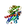 6h17C 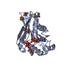 6h35C  6h5bC 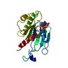 6hjhC  6hjmC  3t1oS S: Starting model for refinement C: citing same article ( |
|---|---|
| Similar structure data |


-Links
 PDBj
PDBj- Assembly
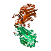
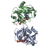
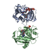
| Deposited unit | 
| ||||||||
|---|---|---|---|---|---|---|---|---|---|
| 1 |
| ||||||||
| Unit cell |
|
-Components
| #1: Protein | Mass: 22042.379 Da / Num. of mol.: 2 Source method: isolated from a genetically manipulated source Source: (gene. exp.) Myxococcus xanthus DK 1622 (bacteria) / Gene: mglA, MXAN_1925 / Production host: Escherichia coli (E. coli) / References: UniProt: Q1DB04#2: Chemical | Guanosine diphosphate  Type: RNA linking / Mass: 443.201 Da / Num. of mol.: 2 / Source method: obtained synthetically / Formula: C10H15N5O11P2 / Comment: GDP, energy-carrying molecule*YM Type: RNA linking / Mass: 443.201 Da / Num. of mol.: 2 / Source method: obtained synthetically / Formula: C10H15N5O11P2 / Comment: GDP, energy-carrying molecule*YM#3: Water | ChemComp-HOH / | Water Mass: 18.015 Da / Num. of mol.: 74 / Source method: isolated from a natural source / Formula: H2O Mass: 18.015 Da / Num. of mol.: 74 / Source method: isolated from a natural source / Formula: H2O |
|---|
-Experimental details
-Experiment
| Experiment | Method: X-RAY DIFFRACTION / Number of used crystals: 1 |
|---|
- Sample preparation
| Crystal | Density Matthews: 3.24 Å3/Da / Density % sol: 61.98 % |
|---|---|
| Crystal grow | Temperature: 293 K / Method: vapor diffusion, hanging drop / pH: 6.5 Details: 0.1M sodium cacodylate pH 6.5, 40% MPD and PEG 8 000 |
-Data collection
| Diffraction | Mean temperature: 100 K |
|---|---|
| Diffraction source | Source: SYNCHROTRON / Site: SOLEIL  / Beamline: PROXIMA 1 / Wavelength: 0.97857 Å / Beamline: PROXIMA 1 / Wavelength: 0.97857 Å |
| Detector | Type: DECTRIS PILATUS 6M / Detector: PIXEL / Date: Jun 27, 2014 |
| Radiation | Protocol: SINGLE WAVELENGTH / Monochromatic (M) / Laue (L): M / Scattering type: x-ray |
| Radiation wavelength | Wavelength: 0.97857 Å / Relative weight: 1 |
| Reflection | Resolution: 1.98→36.13 Å / Num. obs: 40545 / % possible obs: 98 % / Redundancy: 4.4 % / Biso Wilson estimate: 33.56 Å2 / CC1/2: 0.994 / Rmerge(I) obs: 0.1209 / Rrim(I) all: 0.1364 / Net I/σ(I): 13.07 |
| Reflection shell | Resolution: 1.98→2.05 Å / Redundancy: 4.1 % / Rmerge(I) obs: 1.28 / Mean I/σ(I) obs: 0.6 / Num. unique obs: 3634 / CC1/2: 0.744 / Rrim(I) all: 1.276 / % possible all: 100 |
- Processing
| Software |
| ||||||||||||||||||||||||||||||||||||||||||||||||||||||||||||||||||||||||||||||||||||||||||||||||||||||||||||||||||
|---|---|---|---|---|---|---|---|---|---|---|---|---|---|---|---|---|---|---|---|---|---|---|---|---|---|---|---|---|---|---|---|---|---|---|---|---|---|---|---|---|---|---|---|---|---|---|---|---|---|---|---|---|---|---|---|---|---|---|---|---|---|---|---|---|---|---|---|---|---|---|---|---|---|---|---|---|---|---|---|---|---|---|---|---|---|---|---|---|---|---|---|---|---|---|---|---|---|---|---|---|---|---|---|---|---|---|---|---|---|---|---|---|---|---|---|
| Refinement | Method to determine structure: MOLECULAR REPLACEMENT Starting model: 3T1O Resolution: 1.98→36.13 Å / Cor.coef. Fo:Fc: 0.913 / Cor.coef. Fo:Fc free: 0.922 / SU R Cruickshank DPI: 0.185 / Cross valid method: THROUGHOUT / σ(F): 0 / SU R Blow DPI: 0.182 / SU Rfree Blow DPI: 0.152 / SU Rfree Cruickshank DPI: 0.155
| ||||||||||||||||||||||||||||||||||||||||||||||||||||||||||||||||||||||||||||||||||||||||||||||||||||||||||||||||||
| Displacement parameters | Biso mean: 44.6 Å2
| ||||||||||||||||||||||||||||||||||||||||||||||||||||||||||||||||||||||||||||||||||||||||||||||||||||||||||||||||||
| Refine analyze | Luzzati coordinate error obs: 0.33 Å | ||||||||||||||||||||||||||||||||||||||||||||||||||||||||||||||||||||||||||||||||||||||||||||||||||||||||||||||||||
| Refinement step | Cycle: 1 / Resolution: 1.98→36.13 Å
| ||||||||||||||||||||||||||||||||||||||||||||||||||||||||||||||||||||||||||||||||||||||||||||||||||||||||||||||||||
| Refine LS restraints |
| ||||||||||||||||||||||||||||||||||||||||||||||||||||||||||||||||||||||||||||||||||||||||||||||||||||||||||||||||||
| LS refinement shell | Resolution: 1.98→2.05 Å / Total num. of bins used: 50
| ||||||||||||||||||||||||||||||||||||||||||||||||||||||||||||||||||||||||||||||||||||||||||||||||||||||||||||||||||
| Refinement TLS params. | Method: refined / Refine-ID: X-RAY DIFFRACTION
| ||||||||||||||||||||||||||||||||||||||||||||||||||||||||||||||||||||||||||||||||||||||||||||||||||||||||||||||||||
| Refinement TLS group |
|
