 Movie
Movie Controller

[English] 日本語
 Yorodumi
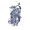
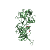
- PDB-6hgd: Crystal structure of Alpha1-antichymotrypsin variant NewBG-0: a n... -
+ Open data

- Basic information
| Entry | Database: PDB / ID: 6hgd | ||||||
|---|---|---|---|---|---|---|---|
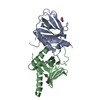
| Title | Crystal structure of Alpha1-antichymotrypsin variant NewBG-0: a new binding globulin variant that is devoid of any cortisol-binding capabilities | ||||||
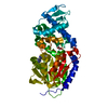
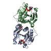
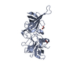
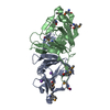
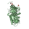
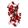
 Components Components | (Alpha-1-antichymotrypsin Alpha 1-antichymotrypsin) x 2 Alpha 1-antichymotrypsin) x 2 | ||||||
 Keywords Keywords | TRANSPORT PROTEIN / Serpin / alpha1-Antichymotrypsin / computational protein design | ||||||
| Function / homology |  Function and homology information Function and homology informationmaintenance of gastrointestinal epithelium / regulation of lipid metabolic process / platelet alpha granule lumen / acute-phase response / serine-type endopeptidase inhibitor activity / azurophil granule lumen / Platelet degranulation / secretory granule lumen / collagen-containing extracellular matrix / blood microparticle ...maintenance of gastrointestinal epithelium / regulation of lipid metabolic process / platelet alpha granule lumen / acute-phase response / serine-type endopeptidase inhibitor activity / azurophil granule lumen / Platelet degranulation / secretory granule lumen / collagen-containing extracellular matrix / blood microparticle / inflammatory response / Neutrophil degranulation / DNA binding / extracellular space / extracellular exosome / extracellular region / nucleusSimilarity search - Function | ||||||
| Biological species |  Homo sapiens (human) Homo sapiens (human) | ||||||
| Method | X-RAY DIFFRACTION / SYNCHROTRON / MOLECULAR REPLACEMENT / Resolution: 1.9 Å | ||||||
 Authors Authors | Gardill, B.R. / Schmidt, K. / Muller, Y.A. | ||||||
 Citation Citation | Journal: J.Struct.Biol. / Year: 2019 Title: NewBG: A surrogate corticosteroid-binding globulin with an unprecedentedly high ligand release efficacy. Authors: Gardill, B.R. / Schmidt, K. / Muller, Y.A. | ||||||
| History |
|
- Structure visualization
| Structure viewer | Molecule: MolmilJmol/JSmol |
|---|
- Downloads & links
-Download
| PDBx/mmCIF format | 6hgd.cif.gz | 98.6 KB | Display | PDBx/mmCIF format |
|---|---|---|---|---|
| PDB format | pdb6hgd.ent.gz | 72.3 KB | Display | PDB format |
| PDBx/mmJSON format | 6hgd.json.gz | Tree view | PDBx/mmJSON format | |
| Others |  Other downloads Other downloads |
-Validation report
| Arichive directory | https://data.pdbj.org/pub/pdb/validation_reports/hg/6hgdftp://data.pdbj.org/pub/pdb/validation_reports/hg/6hgd | HTTPS FTP |
|---|
-Related structure data
| Related structure data |  6hgeC  6hgfC  6hggC  6hghC 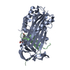 6hgiC  6hgjC  6hgkC  6hglC  6hgmC  6hgnC  1as4S S: Starting model for refinement C: citing same article ( |
|---|---|
| Similar structure data |

-Links
 PDBj
PDBj
- Assembly
| Deposited unit | 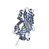
| ||||||||
|---|---|---|---|---|---|---|---|---|---|
| 1 |
| ||||||||
| Unit cell |
|
-Components
| #1: Protein | Alpha 1-antichymotrypsin / ACT / Cell growth-inhibiting gene 24/25 protein / Serpin A3 Mass: 41858.469 Da / Num. of mol.: 1 / Mutation: L24R, E242Q, K244N, K274N, R277G Source method: isolated from a genetically manipulated source Details: - all N-terminal residues that are present in the sample sequence but not in the PDB file could not be modelled due to missing electron density - residues following the sequence ..KITLL are ...Details: - all N-terminal residues that are present in the sample sequence but not in the PDB file could not be modelled due to missing electron density - residues following the sequence ..KITLL are part of chain B, as the protein is a family member of serine proteinase inhibitors (serpins) and proteolytically cleaved between KITLL-SALVE Source: (gene. exp.) Homo sapiens (human) / Gene: SERPINA3, AACT, GIG24, GIG25 / Production host:  Escherichia coli BL21(DE3) (bacteria) / Variant (production host): Star / References: UniProt: P01011 Escherichia coli BL21(DE3) (bacteria) / Variant (production host): Star / References: UniProt: P01011 |
|---|---|
| #2: Protein/peptide | Alpha 1-antichymotrypsin / ACT / Cell growth-inhibiting gene 24/25 protein / Serpin A3 Mass: 4775.638 Da / Num. of mol.: 1 / Mutation: P382D, T383H, D384F, Q386W Source method: isolated from a genetically manipulated source Details: the residues SAL that are present in the sample sequence but not in the PDB file could not be modelled due to missing electron density Source: (gene. exp.) Homo sapiens (human) / Gene: SERPINA3, AACT, GIG24, GIG25 / Production host: Escherichia coli BL21(DE3) (bacteria) / Variant (production host): Star / References: UniProt: P01011 |
| #3: Water | ChemComp-HOH / Water Mass: 18.015 Da / Num. of mol.: 385 / Source method: isolated from a natural source / Formula: H2O Mass: 18.015 Da / Num. of mol.: 385 / Source method: isolated from a natural source / Formula: H2O |
-Experimental details
-Experiment
| Experiment | Method: X-RAY DIFFRACTION / Number of used crystals: 1 |
|---|
- Sample preparation
| Crystal | Density Matthews: 1.92 Å3/Da / Density % sol: 35.87 % |
|---|---|
| Crystal grow | Temperature: 293 K / Method: vapor diffusion, sitting drop Details: 0.2 M magnesium chloride hexahydrate, 0.1 M BIS-Tris pH 6.5, 25 % w/v PEG 3350 |
-Data collection
| Diffraction | Mean temperature: 100 K |
|---|---|
| Diffraction source | Source: SYNCHROTRON / Site: BESSY  / Beamline: 14.1 / Wavelength: 0.91841 Å / Beamline: 14.1 / Wavelength: 0.91841 Å |
| Detector | Type: MARMOSAIC 225 mm CCD / Detector: CCD / Date: Sep 17, 2011 |
| Radiation | Protocol: SINGLE WAVELENGTH / Monochromatic (M) / Laue (L): M / Scattering type: x-ray |
| Radiation wavelength | Wavelength: 0.91841 Å / Relative weight: 1 |
| Reflection | Resolution: 1.9→41.409 Å / Num. obs: 27559 / % possible obs: 99.2 % / Redundancy: 3.73 % / Rrim(I) all: 0.052 / Net I/σ(I): 20.56 |
| Reflection shell | Resolution: 1.9→2.01 Å / Mean I/σ(I) obs: 7.86 / Rrim(I) all: 0.177 |
- Processing
| Software |
| |||||||||||||||||||||||||||||||||||||||||||||||||||||||||||||||||||||||||||||
|---|---|---|---|---|---|---|---|---|---|---|---|---|---|---|---|---|---|---|---|---|---|---|---|---|---|---|---|---|---|---|---|---|---|---|---|---|---|---|---|---|---|---|---|---|---|---|---|---|---|---|---|---|---|---|---|---|---|---|---|---|---|---|---|---|---|---|---|---|---|---|---|---|---|---|---|---|---|---|
| Refinement | Method to determine structure: MOLECULAR REPLACEMENT Starting model: 1AS4 Resolution: 1.9→38.939 Å / SU ML: 0.19 / Cross valid method: FREE R-VALUE / σ(F): 2 / Phase error: 19.23
| |||||||||||||||||||||||||||||||||||||||||||||||||||||||||||||||||||||||||||||
| Solvent computation | Shrinkage radii: 0.9 Å / VDW probe radii: 1.11 Å | |||||||||||||||||||||||||||||||||||||||||||||||||||||||||||||||||||||||||||||
| Refinement step | Cycle: LAST / Resolution: 1.9→38.939 Å
| |||||||||||||||||||||||||||||||||||||||||||||||||||||||||||||||||||||||||||||
| Refine LS restraints |
| |||||||||||||||||||||||||||||||||||||||||||||||||||||||||||||||||||||||||||||
| LS refinement shell |
|
