 Movie
Movie Controller

[English] 日本語
 Yorodumi
- PDB-6hew: Crystal Structure of Ephrin A2 (EphA2) Receptor Protein Kinase wi... -
+ Open data

- Basic information
| Entry | Database: PDB / ID: 6hew | ||||||
|---|---|---|---|---|---|---|---|
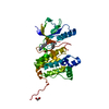
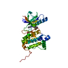
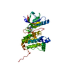
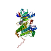
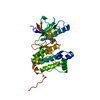
| Title | Crystal Structure of Ephrin A2 (EphA2) Receptor Protein Kinase with the NVP-BHG712 derivative AT069 | ||||||
 Components Components | Ephrin type-A receptor 2 | ||||||
 Keywords Keywords |  TRANSFERASE / Inhibitor / Complex / Protein Tyrosine Kinase TRANSFERASE / Inhibitor / Complex / Protein Tyrosine Kinase | ||||||
| Function / homology |  Function and homology information Function and homology informationnotochord cell development / notochord formation / lens fiber cell morphogenesis / blood vessel endothelial cell proliferation involved in sprouting angiogenesis / negative regulation of lymphangiogenesis / axial mesoderm formation / pericyte cell differentiation / cAMP metabolic process / positive regulation of bicellular tight junction assembly / regulation of blood vessel endothelial cell migration ...notochord cell development / notochord formation / lens fiber cell morphogenesis / blood vessel endothelial cell proliferation involved in sprouting angiogenesis / negative regulation of lymphangiogenesis / axial mesoderm formation / pericyte cell differentiation / cAMP metabolic process / positive regulation of bicellular tight junction assembly / regulation of blood vessel endothelial cell migration / negative regulation of chemokine production / ephrin receptor activity / leading edge membrane / bone remodeling / post-anal tail morphogenesis / response to growth factor / activation of GTPase activity / regulation of lamellipodium assembly / tight junction / branching involved in mammary gland duct morphogenesis / EPH-Ephrin signaling / neural tube development / RND1 GTPase cycle / RND2 GTPase cycle / RND3 GTPase cycle / mammary gland epithelial cell proliferation / RHOV GTPase cycle / EPHA-mediated growth cone collapse / growth factor binding / regulation of cell adhesion mediated by integrin / lamellipodium membrane / RHOU GTPase cycle / RHOG GTPase cycle / EPH-ephrin mediated repulsion of cells / negative regulation of phosphatidylinositol 3-kinase/protein kinase B signal transduction / ephrin receptor signaling pathway / RAC2 GTPase cycle / RAC3 GTPase cycle / vasculogenesis / regulation of angiogenesis / keratinocyte differentiation / RAC1 GTPase cycle / transmembrane receptor protein tyrosine kinase activity / cell chemotaxis / negative regulation of angiogenesis / osteoclast differentiation / regulation of ERK1 and ERK2 cascade / phosphatidylinositol 3-kinase/protein kinase B signal transduction / skeletal system development / molecular function activator activity / cell motility / protein localization to plasma membrane / positive regulation of protein localization to plasma membrane / receptor protein-tyrosine kinase / neuron differentiation / ruffle membrane / osteoblast differentiation / cell surface receptor protein tyrosine kinase signaling pathway / intrinsic apoptotic signaling pathway in response to DNA damage / cell migration / virus receptor activity / lamellipodium / receptor complex / cell adhesion / positive regulation of cell migration / defense response to Gram-positive bacterium / cadherin binding / inflammatory response / phosphorylation / focal adhesion / cell surface / ATP binding / plasma membraneSimilarity search - Function | ||||||
| Biological species |  Homo sapiens (human) Homo sapiens (human) | ||||||
| Method | X-RAY DIFFRACTION / SYNCHROTRON / MOLECULAR REPLACEMENT / Resolution: 1.268 Å | ||||||
 Authors Authors | Kudlinzki, D. / Troester, A. / Witt, K. / Linhard, V.L. / Gande, S.L. / Saxena, K. / Schwalbe, H. | ||||||
 Citation Citation | Journal: To Be Published Title: Effects of NVP-BHG712 chemical modifications on EPHA2 binding and affinity Authors: Troester, A. / Kudlinzki, D. / Saxena, K. / Gande, S. / Schwalbe, H. | ||||||
| History |
|
- Structure visualization
| Structure viewer | Molecule: MolmilJmol/JSmol |
|---|
- Downloads & links
-Download
| PDBx/mmCIF format | 6hew.cif.gz | 131.3 KB | Display | PDBx/mmCIF format |
|---|---|---|---|---|
| PDB format | pdb6hew.ent.gz | 101.4 KB | Display | PDB format |
| PDBx/mmJSON format | 6hew.json.gz | Tree view | PDBx/mmJSON format | |
| Others |  Other downloads Other downloads |
-Validation report
| Arichive directory | https://data.pdbj.org/pub/pdb/validation_reports/he/6hewftp://data.pdbj.org/pub/pdb/validation_reports/he/6hew | HTTPS FTP |
|---|
-Related structure data
| Related structure data |  6hesC  6hetC  6heuC  6hevC  6hexC  6heyC  6q7bC  6q7cC  6q7dC 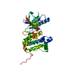 6q7eC 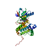 6q7fC 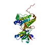 6q7gC 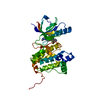 6fnfS S: Starting model for refinement C: citing same article ( |
|---|---|
| Similar structure data |


-Links
 PDBj
PDBj


- Assembly
| Deposited unit | 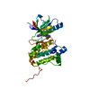
| ||||||||
|---|---|---|---|---|---|---|---|---|---|
| 1 |
| ||||||||
| Unit cell |
|
-Components
| #1: Protein | Mass: 34462.840 Da / Num. of mol.: 1 Source method: isolated from a genetically manipulated source Source: (gene. exp.) Homo sapiens (human) / Gene: EPHA2, ECK / Production host:   Spodoptera frugiperda (fall armyworm) / References: UniProt: P29317 Spodoptera frugiperda (fall armyworm) / References: UniProt: P29317 |
|---|---|
| #2: Chemical | ChemComp-G0E /   Mass: 504.467 Da / Num. of mol.: 1 / Source method: obtained synthetically / Formula: C25H19F3N8O / Feature type: SUBJECT OF INVESTIGATION Mass: 504.467 Da / Num. of mol.: 1 / Source method: obtained synthetically / Formula: C25H19F3N8O / Feature type: SUBJECT OF INVESTIGATION |
| #3: Water | ChemComp-HOH / Water Mass: 18.015 Da / Num. of mol.: 269 / Source method: isolated from a natural source / Formula: H2O Mass: 18.015 Da / Num. of mol.: 269 / Source method: isolated from a natural source / Formula: H2O |
-Experimental details
-Experiment
| Experiment | Method: X-RAY DIFFRACTION / Number of used crystals: 1 |
|---|
- Sample preparation
| Crystal | Density Matthews: 1.96 Å3/Da / Density % sol: 37.24 % |
|---|---|
| Crystal grow | Temperature: 291 K / Method: vapor diffusion, sitting drop / pH: 6 Details: 37.5 % MPD/PEG1000/PEG3350 (MD), 0.1 M Amino Acids Mix (MD), 0.1 M Bis/Tris pH 6.0 |
-Data collection
| Diffraction | Mean temperature: 100 K |
|---|---|
| Diffraction source | Source: SYNCHROTRON / Site: PETRA III, EMBL c/o DESY  / Beamline: P13 (MX1) / Wavelength: 0.97627 Å / Beamline: P13 (MX1) / Wavelength: 0.97627 Å |
| Detector | Type: DECTRIS PILATUS 6M-F / Detector: PIXEL / Date: Dec 20, 2016 |
| Radiation | Protocol: SINGLE WAVELENGTH / Monochromatic (M) / Laue (L): M / Scattering type: x-ray |
| Radiation wavelength | Wavelength: 0.97627 Å / Relative weight: 1 |
| Reflection | Resolution: 1.26→38.42 Å / Num. obs: 69838 / % possible obs: 98.2 % / Redundancy: 6.35 % / Biso Wilson estimate: 21.66 Å2 / CC1/2: 0.996 / Rrim(I) all: 0.203 / Net I/σ(I): 5.86 |
| Reflection shell | Resolution: 1.26→1.34 Å / Redundancy: 5.98 % / Mean I/σ(I) obs: 0.25 / Num. unique obs: 10407 / CC1/2: 0.12 / % possible all: 91 |
- Processing
| Software |
| |||||||||||||||||||||||||||||||||||||||||||||||||||||||||||||||||||||||||||||||||||||||||||||||||||||||||||||||||||||||||||||||||||||||||||||||||||||||||||||||||||||||||||||||
|---|---|---|---|---|---|---|---|---|---|---|---|---|---|---|---|---|---|---|---|---|---|---|---|---|---|---|---|---|---|---|---|---|---|---|---|---|---|---|---|---|---|---|---|---|---|---|---|---|---|---|---|---|---|---|---|---|---|---|---|---|---|---|---|---|---|---|---|---|---|---|---|---|---|---|---|---|---|---|---|---|---|---|---|---|---|---|---|---|---|---|---|---|---|---|---|---|---|---|---|---|---|---|---|---|---|---|---|---|---|---|---|---|---|---|---|---|---|---|---|---|---|---|---|---|---|---|---|---|---|---|---|---|---|---|---|---|---|---|---|---|---|---|---|---|---|---|---|---|---|---|---|---|---|---|---|---|---|---|---|---|---|---|---|---|---|---|---|---|---|---|---|---|---|---|---|---|
| Refinement | Method to determine structure: MOLECULAR REPLACEMENT Starting model: 6FNF Resolution: 1.268→38.418 Å / SU ML: 0.27 / Cross valid method: FREE R-VALUE / σ(F): 1.33 / Phase error: 30.36
| |||||||||||||||||||||||||||||||||||||||||||||||||||||||||||||||||||||||||||||||||||||||||||||||||||||||||||||||||||||||||||||||||||||||||||||||||||||||||||||||||||||||||||||||
| Solvent computation | Shrinkage radii: 0.9 Å / VDW probe radii: 1.11 Å | |||||||||||||||||||||||||||||||||||||||||||||||||||||||||||||||||||||||||||||||||||||||||||||||||||||||||||||||||||||||||||||||||||||||||||||||||||||||||||||||||||||||||||||||
| Refinement step | Cycle: LAST / Resolution: 1.268→38.418 Å
| |||||||||||||||||||||||||||||||||||||||||||||||||||||||||||||||||||||||||||||||||||||||||||||||||||||||||||||||||||||||||||||||||||||||||||||||||||||||||||||||||||||||||||||||
| Refine LS restraints |
| |||||||||||||||||||||||||||||||||||||||||||||||||||||||||||||||||||||||||||||||||||||||||||||||||||||||||||||||||||||||||||||||||||||||||||||||||||||||||||||||||||||||||||||||
| LS refinement shell |
| |||||||||||||||||||||||||||||||||||||||||||||||||||||||||||||||||||||||||||||||||||||||||||||||||||||||||||||||||||||||||||||||||||||||||||||||||||||||||||||||||||||||||||||||
| Refinement TLS params. | Method: refined / Refine-ID: X-RAY DIFFRACTION
| |||||||||||||||||||||||||||||||||||||||||||||||||||||||||||||||||||||||||||||||||||||||||||||||||||||||||||||||||||||||||||||||||||||||||||||||||||||||||||||||||||||||||||||||
| Refinement TLS group |
|
