 Movie
Movie Controller


+ Open data

- Basic information
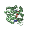
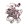
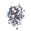
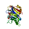
| Entry | Database: PDB / ID: 6hap | ||||||
|---|---|---|---|---|---|---|---|
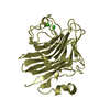
| Title | Adenylate kinase | ||||||
 Components Components | Adenylate kinase | ||||||
 Keywords Keywords | TRANSFERASE / Adenylat kinase | ||||||
| Function / homology |  Function and homology informationadenylate kinase / adenylate kinase activity / AMP salvage / phosphorylation / ATP binding / cytoplasm Function and homology informationadenylate kinase / adenylate kinase activity / AMP salvage / phosphorylation / ATP binding / cytoplasmSimilarity search - Function | ||||||
| Biological species |  Escherichia coli O139:H28 (bacteria) Escherichia coli O139:H28 (bacteria) | ||||||
| Method | X-RAY DIFFRACTION / MOLECULAR REPLACEMENT / Resolution: 2.7 Å | ||||||
 Authors Authors | Kantaev, R. / Inbal, R. / Goldenzweig, A. / Barak, Y. / Dym, O. / Peleg, Y. / Albek, S. / Fleishman, S.J. / Haran, G. | ||||||
 Citation Citation | Journal: J.Phys.Chem.B / Year: 2018 Title: Manipulating the Folding Landscape of a Multidomain Protein. Authors: Kantaev, R. / Riven, I. / Goldenzweig, A. / Barak, Y. / Dym, O. / Peleg, Y. / Albeck, S. / Fleishman, S.J. / Haran, G. | ||||||
| History |
|
- Structure visualization
| Structure viewer | Molecule: MolmilJmol/JSmol |
|---|
- Downloads & links
-Download
| PDBx/mmCIF format | 6hap.cif.gz | 55.3 KB | Display | PDBx/mmCIF format |
|---|---|---|---|---|
| PDB format | pdb6hap.ent.gz | 39.2 KB | Display | PDB format |
| PDBx/mmJSON format | 6hap.json.gz | Tree view | PDBx/mmJSON format | |
| Others |  Other downloads Other downloads |
-Validation report
| Arichive directory | https://data.pdbj.org/pub/pdb/validation_reports/ha/6hapftp://data.pdbj.org/pub/pdb/validation_reports/ha/6hap | HTTPS FTP |
|---|
-Related structure data
| Related structure data | 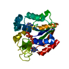 6hamC 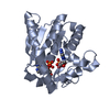 4jzkS S: Starting model for refinement C: citing same article ( |
|---|---|
| Similar structure data |


-Links
 PDBj
PDBj- Assembly
| Deposited unit | 
| ||||||||
|---|---|---|---|---|---|---|---|---|---|
| 1 |
| ||||||||
| Unit cell |
|
-Components
| #1: Protein | / AK / ATP-AMP transphosphorylase / ATP:AMP phosphotransferase / Adenylate monophosphate kinase Mass: 23696.107 Da / Num. of mol.: 1 Source method: isolated from a genetically manipulated source Source: (gene. exp.) Escherichia coli O139:H28 (strain E24377A / ETEC) (bacteria)Strain: E24377A / ETEC / Gene: adk, EcE24377A_0513 Production host: References: UniProt: A7ZIN4, adenylate kinase |
|---|---|
| #2: Chemical | ChemComp-AP5 /   Mass: 916.367 Da / Num. of mol.: 1 / Source method: obtained synthetically / Formula: C20H29N10O22P5 Mass: 916.367 Da / Num. of mol.: 1 / Source method: obtained synthetically / Formula: C20H29N10O22P5 |
-Experimental details
-Experiment
| Experiment | Method: X-RAY DIFFRACTION / Number of used crystals: 1 |
|---|
- Sample preparation
| Crystal | Density Matthews: 2.5 Å3/Da / Density % sol: 50.78 % |
|---|---|
| Crystal grow | Temperature: 298 K / Method: vapor diffusion, hanging drop / pH: 5.5 / Details: 10% PEG 3350 0.05M Tris pH=5.5 |
-Data collection
| Diffraction | Mean temperature: 100 K |
|---|---|
| Diffraction source | Source: ROTATING ANODE / Type: RIGAKU RUH3R / Wavelength: 1.5417 Å |
| Detector | Type: RIGAKU RAXIS IV++ / Detector: IMAGE PLATE / Date: Sep 25, 2017 |
| Radiation | Protocol: SINGLE WAVELENGTH / Monochromatic (M) / Laue (L): M / Scattering type: x-ray |
| Radiation wavelength | Wavelength: 1.5417 Å / Relative weight: 1 |
| Reflection | Resolution: 2.7→43.49 Å / Num. obs: 7058 / % possible obs: 99.7 % / Redundancy: 5.8 % / Rsym value: 0.0356 / Net I/σ(I): 16.48 |
| Reflection shell | Resolution: 2.7→2.8 Å / Redundancy: 5.8 % / Num. unique obs: 696 / Rsym value: 0.3 / % possible all: 99.7 |
- Processing
| Software |
| ||||||||||||||||||||||||
|---|---|---|---|---|---|---|---|---|---|---|---|---|---|---|---|---|---|---|---|---|---|---|---|---|---|
| Refinement | Method to determine structure: MOLECULAR REPLACEMENT Starting model: 4JZK Resolution: 2.7→43.49 Å / SU ML: 0.33 / Cross valid method: FREE R-VALUE / σ(F): 1.35 / Phase error: 33.17
| ||||||||||||||||||||||||
| Solvent computation | Shrinkage radii: 0.9 Å / VDW probe radii: 1.11 Å | ||||||||||||||||||||||||
| Refinement step | Cycle: LAST / Resolution: 2.7→43.49 Å
| ||||||||||||||||||||||||
| Refine LS restraints |
| ||||||||||||||||||||||||
| LS refinement shell |
|
