 Movie
Movie Controller

[English] 日本語
 Yorodumi
- PDB-6gzt: Structure of Chlamydia trachomatis effector protein ChlaDUB1 boun... -
+ Open data

- Basic information
| Entry | Database: PDB / ID: 6gzt | |||||||||
|---|---|---|---|---|---|---|---|---|---|---|
| Title | Structure of Chlamydia trachomatis effector protein ChlaDUB1 bound to Coenzyme A | |||||||||
 Components Components | Deubiquitinase and deneddylase Dub1 | |||||||||
 Keywords Keywords |  HYDROLASE / Enzyme / CE clan / Deubiquitinase / Acetyltransferase HYDROLASE / Enzyme / CE clan / Deubiquitinase / Acetyltransferase | |||||||||
| Function / homology |  Function and homology information Function and homology informationdeNEDDylase activity / deSUMOylase activity / protein desumoylation / protein deneddylation / protein deubiquitination / host cell / cysteine-type deubiquitinase activity / Hydrolases; Acting on peptide bonds (peptidases); Cysteine endopeptidases / proteolysis / extracellular region / membraneSimilarity search - Function | |||||||||
| Biological species |   Chlamydia trachomatis (bacteria) Chlamydia trachomatis (bacteria) | |||||||||
| Method | X-RAY DIFFRACTION / SYNCHROTRON / MOLECULAR REPLACEMENT / Resolution: 2.1 Å | |||||||||
 Authors Authors | Pruneda, J.N. / Komander, D. | |||||||||
| Funding support |  United Kingdom, 2items United Kingdom, 2items
| |||||||||
 Citation Citation | Journal: Nat Microbiol / Year: 2018 Title: A Chlamydia effector combining deubiquitination and acetylation activities induces Golgi fragmentation. Authors: Pruneda, J.N. / Bastidas, R.J. / Bertsoulaki, E. / Swatek, K.N. / Santhanam, B. / Clague, M.J. / Valdivia, R.H. / Urbe, S. / Komander, D. | |||||||||
| History |
|
- Structure visualization
| Structure viewer | Molecule: MolmilJmol/JSmol |
|---|
- Downloads & links
-Download
| PDBx/mmCIF format | 6gzt.cif.gz | 74.7 KB | Display | PDBx/mmCIF format |
|---|---|---|---|---|
| PDB format | pdb6gzt.ent.gz | 53.5 KB | Display | PDB format |
| PDBx/mmJSON format | 6gzt.json.gz | Tree view | PDBx/mmJSON format | |
| Others |  Other downloads Other downloads |
-Validation report
| Arichive directory | https://data.pdbj.org/pub/pdb/validation_reports/gz/6gztftp://data.pdbj.org/pub/pdb/validation_reports/gz/6gzt | HTTPS FTP |
|---|
-Related structure data
| Related structure data | 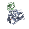 6gzsC 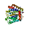 6gzuC 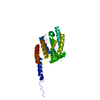 5hagS S: Starting model for refinement C: citing same article ( |
|---|---|
| Similar structure data |


-Links
 PDBj
PDBj


- Assembly
| Deposited unit | 
| ||||||||
|---|---|---|---|---|---|---|---|---|---|
| 1 |
| ||||||||
| Unit cell |
| ||||||||
| Components on special symmetry positions |
|
-Components
| #1: Protein | Mass: 31307.244 Da / Num. of mol.: 1 / Fragment: UNP residues 130-401 Source method: isolated from a genetically manipulated source Source: (gene. exp.) Chlamydia trachomatis (bacteria) / Gene: cdu1, CTL0247 / Production host: Escherichia coli (E. coli)References: UniProt: B0B9A0, Hydrolases; Acting on peptide bonds (peptidases); Cysteine endopeptidases | ||||||
|---|---|---|---|---|---|---|---|
| #2: Chemical | ChemComp-GOL / Glycerol  Mass: 92.094 Da / Num. of mol.: 4 / Source method: obtained synthetically / Formula: C3H8O3 Mass: 92.094 Da / Num. of mol.: 4 / Source method: obtained synthetically / Formula: C3H8O3#3: Chemical | ChemComp-SO4 / | Sulfate  Mass: 96.063 Da / Num. of mol.: 1 / Source method: obtained synthetically / Formula: SO4 Mass: 96.063 Da / Num. of mol.: 1 / Source method: obtained synthetically / Formula: SO4#4: Chemical | ChemComp-COA / | Coenzyme A  Mass: 767.534 Da / Num. of mol.: 1 / Source method: obtained synthetically / Formula: C21H36N7O16P3S Mass: 767.534 Da / Num. of mol.: 1 / Source method: obtained synthetically / Formula: C21H36N7O16P3S#5: Water | ChemComp-HOH / | Water Mass: 18.015 Da / Num. of mol.: 147 / Source method: isolated from a natural source / Formula: H2O Mass: 18.015 Da / Num. of mol.: 147 / Source method: isolated from a natural source / Formula: H2O |
-Experimental details
-Experiment
| Experiment | Method: X-RAY DIFFRACTION / Number of used crystals: 1 |
|---|
- Sample preparation
| Crystal | Density Matthews: 3.11 Å3/Da / Density % sol: 60.39 % |
|---|---|
| Crystal grow | Temperature: 291 K / Method: vapor diffusion, sitting drop / Details: 0.1 M HEPES (pH 7.2), 20% PEG 8000 |
-Data collection
| Diffraction | Mean temperature: 100 K |
|---|---|
| Diffraction source | Source: SYNCHROTRON / Site: Diamond / Beamline: I04 / Wavelength: 0.9795 Å |
| Detector | Type: DECTRIS PILATUS 6M-F / Detector: PIXEL / Date: May 28, 2015 |
| Radiation | Protocol: SINGLE WAVELENGTH / Monochromatic (M) / Laue (L): M / Scattering type: x-ray |
| Radiation wavelength | Wavelength: 0.9795 Å / Relative weight: 1 |
| Reflection | Resolution: 2.1→66.32 Å / Num. obs: 22779 / % possible obs: 99.9 % / Redundancy: 5.1 % / Rmerge(I) obs: 0.085 / Net I/σ(I): 10.3 |
| Reflection shell | Resolution: 2.1→2.16 Å / Redundancy: 5.2 % / Rmerge(I) obs: 0.653 / Mean I/σ(I) obs: 2.1 / % possible all: 100 |
- Processing
| Software |
| |||||||||||||||||||||||||||||||||||||||||||||||||||||||||||||||
|---|---|---|---|---|---|---|---|---|---|---|---|---|---|---|---|---|---|---|---|---|---|---|---|---|---|---|---|---|---|---|---|---|---|---|---|---|---|---|---|---|---|---|---|---|---|---|---|---|---|---|---|---|---|---|---|---|---|---|---|---|---|---|---|---|
| Refinement | Method to determine structure: MOLECULAR REPLACEMENT Starting model: 5HAG Resolution: 2.1→46.894 Å / SU ML: 0.25 / Cross valid method: FREE R-VALUE / σ(F): 1.35 / Phase error: 23.08
| |||||||||||||||||||||||||||||||||||||||||||||||||||||||||||||||
| Solvent computation | Shrinkage radii: 0.9 Å / VDW probe radii: 1.11 Å | |||||||||||||||||||||||||||||||||||||||||||||||||||||||||||||||
| Refinement step | Cycle: LAST / Resolution: 2.1→46.894 Å
| |||||||||||||||||||||||||||||||||||||||||||||||||||||||||||||||
| Refine LS restraints |
| |||||||||||||||||||||||||||||||||||||||||||||||||||||||||||||||
| LS refinement shell |
|
