 Movie
Movie Controller


+ Open data

- Basic information
| Entry | Database: PDB / ID: 6gu8 | |||||||||
|---|---|---|---|---|---|---|---|---|---|---|
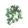
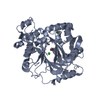
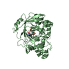
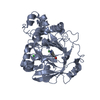
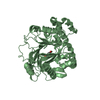
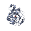
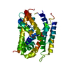
| Title | Glucuronoyl Esterase from Solibacter usitatus | |||||||||
 Components Components | Putative acetyl xylan esterase | |||||||||
 Keywords Keywords |  HYDROLASE / Carbohydrate Esterase HYDROLASE / Carbohydrate Esterase | |||||||||
| Function / homology | Alpha/Beta hydrolase fold / DI(HYDROXYETHYL)ETHER / Putative acetyl xylan esterase Function and homology information Function and homology information | |||||||||
| Biological species |  Candidatus Solibacter usitatus Ellin6076 (bacteria) Candidatus Solibacter usitatus Ellin6076 (bacteria) | |||||||||
| Method | X-RAY DIFFRACTION / SYNCHROTRON / SAD / Resolution: 2.01807823457 Å | |||||||||
 Authors Authors | Lo Leggio, L. / Larsbrink, J. / Meland Knudsen, R. / Mazurkewich, S. / Navarro Poulsen, J.C. | |||||||||
| Funding support |  Denmark, 2items Denmark, 2items
| |||||||||
 Citation Citation | Journal: Biotechnol Biofuels / Year: 2018 Title: Biochemical and structural features of diverse bacterial glucuronoyl esterases facilitating recalcitrant biomass conversion. Authors: Arnling Baath, J. / Mazurkewich, S. / Knudsen, R.M. / Poulsen, J.N. / Olsson, L. / Lo Leggio, L. / Larsbrink, J. | |||||||||
| History |
|
- Structure visualization
| Structure viewer | Molecule: MolmilJmol/JSmol |
|---|
- Downloads & links
-Download
| PDBx/mmCIF format | 6gu8.cif.gz | 200.7 KB | Display | PDBx/mmCIF format |
|---|---|---|---|---|
| PDB format | pdb6gu8.ent.gz | 138.4 KB | Display | PDB format |
| PDBx/mmJSON format | 6gu8.json.gz | Tree view | PDBx/mmJSON format | |
| Others |  Other downloads Other downloads |
-Validation report
| Arichive directory | https://data.pdbj.org/pub/pdb/validation_reports/gu/6gu8ftp://data.pdbj.org/pub/pdb/validation_reports/gu/6gu8 | HTTPS FTP |
|---|
-Related structure data


-Links
 PDBj
PDBj- Assembly
| Deposited unit | 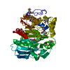
| ||||||||||||
|---|---|---|---|---|---|---|---|---|---|---|---|---|---|
| 1 |
| ||||||||||||
| Unit cell |
|
-Components
| #1: Protein | Mass: 44213.602 Da / Num. of mol.: 1 Source method: isolated from a genetically manipulated source Source: (gene. exp.) Candidatus Solibacter usitatus Ellin6076 (bacteria)Gene: Acid_4275 / Production host: Escherichia coli BL21(DE3) (bacteria) / References: UniProt: Q01YM8 | ||||
|---|---|---|---|---|---|
| #2: Chemical | ChemComp-EDO / Ethylene glycol  Mass: 62.068 Da / Num. of mol.: 8 / Source method: obtained synthetically / Formula: C2H6O2 Mass: 62.068 Da / Num. of mol.: 8 / Source method: obtained synthetically / Formula: C2H6O2#3: Chemical | Diethylene glycol  Mass: 106.120 Da / Num. of mol.: 3 / Source method: obtained synthetically / Formula: C4H10O3 Mass: 106.120 Da / Num. of mol.: 3 / Source method: obtained synthetically / Formula: C4H10O3#4: Water | ChemComp-HOH / | Water Mass: 18.015 Da / Num. of mol.: 277 / Source method: isolated from a natural source / Formula: H2O Mass: 18.015 Da / Num. of mol.: 277 / Source method: isolated from a natural source / Formula: H2O |
-Experimental details
-Experiment
| Experiment | Method: X-RAY DIFFRACTION / Number of used crystals: 1 |
|---|
- Sample preparation
| Crystal | Density Matthews: 2.42 Å3/Da / Density % sol: 49.2 % |
|---|---|
| Crystal grow | Temperature: 293.15 K / Method: vapor diffusion, sitting drop / pH: 8 Details: Reservoir composition: 0.02 M D-glucose, 0.02 M D-mannose, 0.02 M D-galactose, 0.02 M L-fucose, 0.02 M D-xylose, 0.02 M N-acetyl-D-glucosamine, 0.05 M Tris, 0.05 M BICINE, 20% v/v PEG500MME, ...Details: Reservoir composition: 0.02 M D-glucose, 0.02 M D-mannose, 0.02 M D-galactose, 0.02 M L-fucose, 0.02 M D-xylose, 0.02 M N-acetyl-D-glucosamine, 0.05 M Tris, 0.05 M BICINE, 20% v/v PEG500MME, 10% w/v PEG20000 Drop size and composition: Sitting drops of 0.3 ul were mixed in a protein:reservoir volume ratio of 1:1 using 23.6 mg/mL of SuCE15C-SeMet in 20 mM TRIS pH 8.0 |
-Data collection
| Diffraction | Mean temperature: 100 K |
|---|---|
| Diffraction source | Source: SYNCHROTRON / Site: PETRA III, DESY  / Beamline: P11 / Wavelength: 0.979 Å / Beamline: P11 / Wavelength: 0.979 Å |
| Detector | Type: DECTRIS PILATUS 6M / Detector: PIXEL / Date: Aug 26, 2017 |
| Radiation | Protocol: SINGLE WAVELENGTH / Monochromatic (M) / Laue (L): M / Scattering type: x-ray |
| Radiation wavelength | Wavelength: 0.979 Å / Relative weight: 1 |
| Reflection | Resolution: 2.018→47.29 Å / Num. obs: 59163 / % possible obs: 99.95 % / Redundancy: 2 % / Biso Wilson estimate: 32.6808653387 Å2 / CC1/2: 0.999 / Rmerge(I) obs: 0.0691 / Net I/σ(I): 9.27 |
| Reflection shell | Resolution: 2.02→2.09 Å / Redundancy: 2 % / Rmerge(I) obs: 0.4261 / Mean I/σ(I) obs: 1.35 / Num. unique obs: 5723 / CC1/2: 0.889 / % possible all: 99.65 |
- Processing
| Software |
| ||||||||||||||||||||||||||||||||||||||||||||||||||||||||
|---|---|---|---|---|---|---|---|---|---|---|---|---|---|---|---|---|---|---|---|---|---|---|---|---|---|---|---|---|---|---|---|---|---|---|---|---|---|---|---|---|---|---|---|---|---|---|---|---|---|---|---|---|---|---|---|---|---|
| Refinement | Method to determine structure: SAD / Resolution: 2.01807823457→47.2880572634 Å / SU ML: 0.302442641961 / Cross valid method: FREE R-VALUE / σ(F): 1.35372315283 / Phase error: 27.8375960532
| ||||||||||||||||||||||||||||||||||||||||||||||||||||||||
| Solvent computation | Shrinkage radii: 0.9 Å / VDW probe radii: 1.11 Å | ||||||||||||||||||||||||||||||||||||||||||||||||||||||||
| Displacement parameters | Biso mean: 45.4221082505 Å2 | ||||||||||||||||||||||||||||||||||||||||||||||||||||||||
| Refinement step | Cycle: LAST / Resolution: 2.01807823457→47.2880572634 Å
| ||||||||||||||||||||||||||||||||||||||||||||||||||||||||
| Refine LS restraints |
| ||||||||||||||||||||||||||||||||||||||||||||||||||||||||
| LS refinement shell |
|
