 Movie
Movie Controller


+ Open data

- Basic information
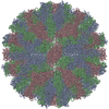
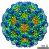
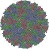
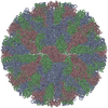
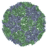
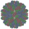
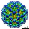
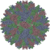
| Entry | Database: PDB / ID: 6gsh | |||||||||
|---|---|---|---|---|---|---|---|---|---|---|
| Title | Feline Calicivirus Strain F9 | |||||||||
 Components Components | VP1 | |||||||||
 Keywords Keywords |  VIRUS / Capsid / Calicivirus / Vesivirus / Vp1 VIRUS / Capsid / Calicivirus / Vesivirus / Vp1 | |||||||||
| Function / homology |  Function and homology information Function and homology information | |||||||||
| Biological species |  Feline calicivirus Feline calicivirus | |||||||||
| Method | ELECTRON MICROSCOPY / single particle reconstruction / cryo EM / Resolution: 3 Å | |||||||||
 Authors Authors | Conley, M.J. / Bhella, D. | |||||||||
| Funding support |  United Kingdom, 2items United Kingdom, 2items
| |||||||||
 Citation Citation | Journal: Nature / Year: 2019 Title: Calicivirus VP2 forms a portal-like assembly following receptor engagement. Authors: Michaela J Conley / Marion McElwee / Liyana Azmi / Mads Gabrielsen / Olwyn Byron / Ian G Goodfellow / David Bhella / Abstract: To initiate infection, many viruses enter their host cells by triggering endocytosis following receptor engagement. However, the mechanisms by which non-enveloped viruses escape the endosome are ...To initiate infection, many viruses enter their host cells by triggering endocytosis following receptor engagement. However, the mechanisms by which non-enveloped viruses escape the endosome are poorly understood. Here we present near-atomic-resolution cryo-electron microscopy structures for feline calicivirus both undecorated and labelled with a soluble fragment of its cellular receptor, feline junctional adhesion molecule A. We show that VP2, a minor capsid protein encoded by all caliciviruses, forms a large portal-like assembly at a unique three-fold axis of symmetry, following receptor engagement. This assembly-which was not detected in undecorated virions-is formed of twelve copies of VP2, arranged with their hydrophobic N termini pointing away from the virion surface. Local rearrangement at the portal site leads to the opening of a pore in the capsid shell. We hypothesize that the portal-like assembly functions as a channel for the delivery of the calicivirus genome, through the endosomal membrane, into the cytoplasm of a host cell, thereby initiating infection. VP2 was previously known to be critical for the production of infectious virus; our findings provide insights into its structure and function that advance our understanding of the Caliciviridae. | |||||||||
| History |
|
- Structure visualization
| Movie |
Movie viewer |
|---|---|
| Structure viewer | Molecule: MolmilJmol/JSmol |
- Downloads & links
-Download
| PDBx/mmCIF format | 6gsh.cif.gz | 503.5 KB | Display | PDBx/mmCIF format |
|---|---|---|---|---|
| PDB format | pdb6gsh.ent.gz | 434.3 KB | Display | PDB format |
| PDBx/mmJSON format | 6gsh.json.gz | Tree view | PDBx/mmJSON format | |
| Others |  Other downloads Other downloads |
-Validation report
| Arichive directory | https://data.pdbj.org/pub/pdb/validation_reports/gs/6gshftp://data.pdbj.org/pub/pdb/validation_reports/gs/6gsh | HTTPS FTP |
|---|
-Related structure data
| Related structure data | 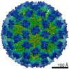 0054MUC 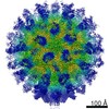 0056C 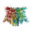 6gsiC M: map data used to model this data U: unfit; in different coordinate system*YM C: citing same article ( |
|---|---|
| Similar structure data | |
| EM raw data | EMPIAR-10192 (Title: Calicivirus VP2 forms a portal to mediate endosome escape Data size: 324.9 Data #1: Motion corrected micrographs of feline calicivirus strain F9 [micrographs - single frame]) |
-Links
 PDBj
PDBj

- Assembly
| Deposited unit | 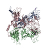
|
|---|---|
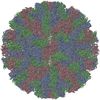
| 1 | x 60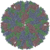
|
-Components
| #1: Protein | Mass: 73346.664 Da / Num. of mol.: 3 Source method: isolated from a genetically manipulated source Source: (gene. exp.) Feline calicivirus / Cell (production host): Crandell Reese Feline Kidney cellsCell line (production host): Crandell Reese Feline Kidney cells Production host:  Felis catus (domestic cat) / References: UniProt: A2T4P8 Felis catus (domestic cat) / References: UniProt: A2T4P8#2: Chemical |   Mass: 39.098 Da / Num. of mol.: 3 / Source method: obtained synthetically / Formula: K Mass: 39.098 Da / Num. of mol.: 3 / Source method: obtained synthetically / Formula: K |
|---|
-Experimental details
-Experiment
| Experiment | Method: ELECTRON MICROSCOPY |
|---|---|
| EM experiment | Aggregation state: PARTICLE / 3D reconstruction method: single particle reconstruction |
- Sample preparation
| Component | Name: T=3 Icosahedral Capsid. / Type: COMPLEX / Details: T=3 Icosahedral Capsid / Entity ID: #1 / Source: RECOMBINANT |
|---|---|
| Molecular weight | Experimental value: NO |
| Source (natural) | Organism: Feline calicivirus |
| Source (recombinant) | Organism: Felis catus (domestic cat) |
| Details of virus | Empty: NO / Enveloped: NO / Isolate: STRAIN / Type: VIRION |
| Natural host | Organism: Felis catus |
| Virus shell | Name: Capsid / Diameter: 400 nm / Triangulation number (T number): 3 |
| Buffer solution | pH: 7.2 / Details: Phosphate buffered saline |
| Specimen | Conc.: 1 mg/ml / Embedding applied: NO / Shadowing applied: NO / Staining applied: NO / Vitrification applied: YES / Details: Purified enveloped virions |
| Vitrification | Instrument: FEI VITROBOT MARK IV / Cryogen name: ETHANE / Humidity: 100 % / Chamber temperature: 277 K |
- Electron microscopy imaging
| Experimental equipment |  Model: Titan Krios / Image courtesy: FEI Company |
|---|---|
| Microscopy | Model: FEI TITAN KRIOS |
| Electron gun | Electron source: FIELD EMISSION GUN / Accelerating voltage: 300 kV / Illumination mode: FLOOD BEAM |
| Electron lens | Mode: BRIGHT FIELDBright-field microscopy / Nominal magnification: 75000 X / Cs: 2.7 mm |
| Image recording | Electron dose: 63 e/Å2 / Detector mode: INTEGRATING / Film or detector model: FEI FALCON III (4k x 4k) / Num. of grids imaged: 1 / Num. of real images: 5198 Details: Each micrograph was recorded as a movie of 50 individual fractions with a total dose of 63 e/angstrom squared |
- Processing
| Software | Name: PHENIX / Version: 1.13_2998: / Classification: refinement | ||||||||||||||||||||||||||||
|---|---|---|---|---|---|---|---|---|---|---|---|---|---|---|---|---|---|---|---|---|---|---|---|---|---|---|---|---|---|
| EM software |
| ||||||||||||||||||||||||||||
| Image processing | Details: Images were motion-corrected using motioncor2 Defocus estimation was performed using GCTF | ||||||||||||||||||||||||||||
| CTF correction | Details: CTF correction was implemented through Relion / Type: PHASE FLIPPING AND AMPLITUDE CORRECTION | ||||||||||||||||||||||||||||
| Particle selection | Num. of particles selected: 59531 / Details: Autopicking in Relion | ||||||||||||||||||||||||||||
| Symmetry | Point symmetry: I (icosahedral) | ||||||||||||||||||||||||||||
| 3D reconstruction | Resolution: 3 Å / Resolution method: FSC 0.143 CUT-OFF / Num. of particles: 41436 / Symmetry type: POINT |
