 Movie
Movie Controller

[English] 日本語
 Yorodumi
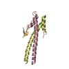
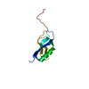
- PDB-6gp7: Cell division regulator, B. subtilis GpsB, in complex with peptid... -
+ Open data

- Basic information
| Entry | Database: PDB / ID: 6gp7 | ||||||
|---|---|---|---|---|---|---|---|
| Title | Cell division regulator, B. subtilis GpsB, in complex with peptide fragment of Penicillin Binding Protein PBP1A | ||||||
 Components Components |
| ||||||
 Keywords Keywords |  CELL CYCLE / Bacterial cell division regulator / peptidoglycan synthesis regulator / penicillin binding protein interaction partner / protein-peptide complex CELL CYCLE / Bacterial cell division regulator / peptidoglycan synthesis regulator / penicillin binding protein interaction partner / protein-peptide complex | ||||||
| Function / homology | Cell cycle protein GpsB / DivIVA family / DivIVA domain / DivIVA protein / regulation of cell shape / cell cycle / cell division / cytoplasm / Cell cycle protein GpsB Function and homology information Function and homology information | ||||||
| Biological species |  Bacillus subtilis subsp. subtilis str. 168 (bacteria) Bacillus subtilis subsp. subtilis str. 168 (bacteria) | ||||||
| Method | X-RAY DIFFRACTION / SYNCHROTRON / MOLECULAR REPLACEMENT / Resolution: 1.95001799213 Å | ||||||
 Authors Authors | Cleverley, R.M. / Lewis, R.J. | ||||||
| Funding support |  United Kingdom, 1items United Kingdom, 1items
| ||||||
 Citation Citation | Journal: Nat Commun / Year: 2019 Title: The cell cycle regulator GpsB functions as cytosolic adaptor for multiple cell wall enzymes. Authors: Cleverley, R.M. / Rutter, Z.J. / Rismondo, J. / Corona, F. / Tsui, H.T. / Alatawi, F.A. / Daniel, R.A. / Halbedel, S. / Massidda, O. / Winkler, M.E. / Lewis, R.J. | ||||||
| History |
|
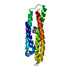
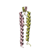
- Structure visualization
| Structure viewer | Molecule: MolmilJmol/JSmol |
|---|
- Downloads & links
-Download
| PDBx/mmCIF format | 6gp7.cif.gz | 80.9 KB | Display | PDBx/mmCIF format |
|---|---|---|---|---|
| PDB format | pdb6gp7.ent.gz | 50 KB | Display | PDB format |
| PDBx/mmJSON format | 6gp7.json.gz | Tree view | PDBx/mmJSON format | |
| Others |  Other downloads Other downloads |
-Validation report
| Arichive directory | https://data.pdbj.org/pub/pdb/validation_reports/gp/6gp7ftp://data.pdbj.org/pub/pdb/validation_reports/gp/6gp7 | HTTPS FTP |
|---|
-Related structure data
| Related structure data | 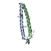 6gpzC  6gqaC 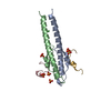 6gqnC  4ug3S S: Starting model for refinement C: citing same article ( |
|---|---|
| Similar structure data |


-Links
 PDBj
PDBj- Assembly
| Deposited unit | 
| ||||||||||||
|---|---|---|---|---|---|---|---|---|---|---|---|---|---|
| 1 |
| ||||||||||||
| Unit cell |
|
-Components
| #1: Protein | / Guiding PBP1-shuttling protein Mass: 7610.684 Da / Num. of mol.: 2 Source method: isolated from a genetically manipulated source Source: (gene. exp.) Bacillus subtilis subsp. subtilis str. 168 Gene: gpsB, ypsB, BSU22180 / Production host: Escherichia coli BL21(DE3) / References: UniProt: P0CI74 #2: Protein/peptide | | Mass: 2030.251 Da / Num. of mol.: 1 / Source method: obtained synthetically Source: (synth.) Bacillus subtilis subsp. subtilis str. 168 (bacteria)#3: Chemical | ChemComp-MG / |   Mass: 24.305 Da / Num. of mol.: 1 / Source method: obtained synthetically / Formula: Mg Mass: 24.305 Da / Num. of mol.: 1 / Source method: obtained synthetically / Formula: Mg#4: Water | ChemComp-HOH / | Water Mass: 18.015 Da / Num. of mol.: 74 / Source method: isolated from a natural source / Formula: H2O Mass: 18.015 Da / Num. of mol.: 74 / Source method: isolated from a natural source / Formula: H2O |
|---|
-Experimental details
-Experiment
| Experiment | Method: X-RAY DIFFRACTION / Number of used crystals: 1 |
|---|
- Sample preparation
| Crystal | Density Matthews: 2.34 Å3/Da / Density % sol: 47.42 % |
|---|---|
| Crystal grow | Temperature: 295 K / Method: vapor diffusion / pH: 7.5 Details: 0.1 M HEPES/MOPS pH 7.5, 12.5% MPD, 12.5 % PEG 1000 12.5% PEG 3350, 0.03M MgCl2, 0.03M CaCl2 |
-Data collection
| Diffraction | Mean temperature: 100 K |
|---|---|
| Diffraction source | Source: SYNCHROTRON / Site: Diamond / Beamline: I24 / Wavelength: 0.9686 Å |
| Detector | Type: DECTRIS PILATUS3 S 6M / Detector: PIXEL / Date: Feb 13, 1996 |
| Radiation | Protocol: SINGLE WAVELENGTH / Monochromatic (M) / Laue (L): M / Scattering type: x-ray |
| Radiation wavelength | Wavelength: 0.9686 Å / Relative weight: 1 |
| Reflection | Resolution: 1.95→45.6 Å / Num. obs: 11206 / % possible obs: 100 % / Redundancy: 7 % / Biso Wilson estimate: 24.8484214955 Å2 / CC1/2: 0.994 / Rpim(I) all: 0.059 / Net I/σ(I): 8.8 |
| Reflection shell | Resolution: 1.95→2 Å / Redundancy: 7.1 % / Mean I/σ(I) obs: 2.1 / Num. unique obs: 768 / CC1/2: 0.72 / Rpim(I) all: 0.473 / % possible all: 100 |
- Processing
| Software |
| |||||||||||||||||||||||||||||||||||||||||||||||||||||||||||||||||||||||||||||||||||||||||||||||||||||||||||||||||||||||||||||||||||||||||||||||||||||||||||||||||||||||||||||||
|---|---|---|---|---|---|---|---|---|---|---|---|---|---|---|---|---|---|---|---|---|---|---|---|---|---|---|---|---|---|---|---|---|---|---|---|---|---|---|---|---|---|---|---|---|---|---|---|---|---|---|---|---|---|---|---|---|---|---|---|---|---|---|---|---|---|---|---|---|---|---|---|---|---|---|---|---|---|---|---|---|---|---|---|---|---|---|---|---|---|---|---|---|---|---|---|---|---|---|---|---|---|---|---|---|---|---|---|---|---|---|---|---|---|---|---|---|---|---|---|---|---|---|---|---|---|---|---|---|---|---|---|---|---|---|---|---|---|---|---|---|---|---|---|---|---|---|---|---|---|---|---|---|---|---|---|---|---|---|---|---|---|---|---|---|---|---|---|---|---|---|---|---|---|---|---|---|
| Refinement | Method to determine structure: MOLECULAR REPLACEMENT Starting model: 4UG3 Resolution: 1.95001799213→45.5686210377 Å / SU ML: 0.161620864616 / Cross valid method: FREE R-VALUE / σ(F): 1.37278577122 / Phase error: 22.7922494119
| |||||||||||||||||||||||||||||||||||||||||||||||||||||||||||||||||||||||||||||||||||||||||||||||||||||||||||||||||||||||||||||||||||||||||||||||||||||||||||||||||||||||||||||||
| Solvent computation | Shrinkage radii: 0.9 Å / VDW probe radii: 1.11 Å | |||||||||||||||||||||||||||||||||||||||||||||||||||||||||||||||||||||||||||||||||||||||||||||||||||||||||||||||||||||||||||||||||||||||||||||||||||||||||||||||||||||||||||||||
| Displacement parameters | Biso mean: 34.5778482729 Å2 | |||||||||||||||||||||||||||||||||||||||||||||||||||||||||||||||||||||||||||||||||||||||||||||||||||||||||||||||||||||||||||||||||||||||||||||||||||||||||||||||||||||||||||||||
| Refinement step | Cycle: LAST / Resolution: 1.95001799213→45.5686210377 Å
| |||||||||||||||||||||||||||||||||||||||||||||||||||||||||||||||||||||||||||||||||||||||||||||||||||||||||||||||||||||||||||||||||||||||||||||||||||||||||||||||||||||||||||||||
| Refine LS restraints |
| |||||||||||||||||||||||||||||||||||||||||||||||||||||||||||||||||||||||||||||||||||||||||||||||||||||||||||||||||||||||||||||||||||||||||||||||||||||||||||||||||||||||||||||||
| LS refinement shell |
| |||||||||||||||||||||||||||||||||||||||||||||||||||||||||||||||||||||||||||||||||||||||||||||||||||||||||||||||||||||||||||||||||||||||||||||||||||||||||||||||||||||||||||||||
| Refinement TLS params. | Method: refined / Refine-ID: X-RAY DIFFRACTION
| |||||||||||||||||||||||||||||||||||||||||||||||||||||||||||||||||||||||||||||||||||||||||||||||||||||||||||||||||||||||||||||||||||||||||||||||||||||||||||||||||||||||||||||||
| Refinement TLS group |
|
