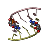
 Movie
Movie Controller

[English] 日本語
 Yorodumi
- PDB-6gn2: Racemic crystal structure of A-DNA duplex formed from d(CCCGGG) i... -
+ Open data

- Basic information
| Entry | Database: PDB / ID: 6gn2 | ||||||||||||||||||||||||||||
|---|---|---|---|---|---|---|---|---|---|---|---|---|---|---|---|---|---|---|---|---|---|---|---|---|---|---|---|---|---|
| Title | Racemic crystal structure of A-DNA duplex formed from d(CCCGGG) in space group R3 | ||||||||||||||||||||||||||||
 Components Components | DNA (5'-D(* Keywords Keywords DNA / Racemate DNA / RacemateFunction / homology | trimethylamine oxide / DNA Function and homology information Function and homology informationBiological species | synthetic construct (others) | Method | X-RAY DIFFRACTION / MOLECULAR REPLACEMENT / Resolution: 2.48 Å  Authors AuthorsMandal, P.K. / Collie, G.W. / Kauffmann, B. / Huc, I. | Funding support | |  France, 1items France, 1items
 CitationJournal: Acta Crystallogr D Struct Biol / Year: 2022 CitationJournal: Acta Crystallogr D Struct Biol / Year: 2022Title: Racemic crystal structures of A-DNA duplexes. Authors: Mandal, P.K. / Collie, G.W. / Kauffmann, B. / Huc, I. #1: Journal: Angew. Chem. Int. Ed. / Year: 2014Title: Racemic DNA crystallography Authors: Mandal, P.K. / Collie, G.W. / Kauffmann, B. / Huc, I. History |
|
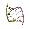
- Structure visualization
| Structure viewer | Molecule: MolmilJmol/JSmol |
|---|
- Downloads & links
-Download
| PDBx/mmCIF format | 6gn2.cif.gz | 35.6 KB | Display | PDBx/mmCIF format |
|---|---|---|---|---|
| PDB format | pdb6gn2.ent.gz | 25.9 KB | Display | PDB format |
| PDBx/mmJSON format | 6gn2.json.gz | Tree view | PDBx/mmJSON format | |
| Others |  Other downloads Other downloads |
-Validation report
| Arichive directory | https://data.pdbj.org/pub/pdb/validation_reports/gn/6gn2ftp://data.pdbj.org/pub/pdb/validation_reports/gn/6gn2 | HTTPS FTP |
|---|
-Related structure data
| Related structure data |  6gn3C  1vt5S S: Starting model for refinement C: citing same article ( |
|---|---|
| Similar structure data |


-Links
 PDBj
PDBj

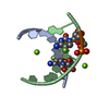
- Assembly
| Deposited unit | 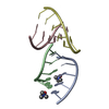
| ||||||||
|---|---|---|---|---|---|---|---|---|---|
| 1 | 
| ||||||||
| 2 | 
| ||||||||
| Unit cell |
|
-Components
| #1: DNA chain | Mass: 1810.205 Da / Num. of mol.: 4 / Source method: obtained synthetically / Source: (synth.) synthetic construct (others) #2: Chemical | Trimethylamine N-oxide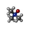  Mass: 75.110 Da / Num. of mol.: 2 / Source method: obtained synthetically / Formula: C3H9NO Mass: 75.110 Da / Num. of mol.: 2 / Source method: obtained synthetically / Formula: C3H9NO#3: Water | ChemComp-HOH / | Water Mass: 18.015 Da / Num. of mol.: 49 / Source method: isolated from a natural source / Formula: H2O Mass: 18.015 Da / Num. of mol.: 49 / Source method: isolated from a natural source / Formula: H2O |
|---|
-Experimental details
-Experiment
| Experiment | Method: X-RAY DIFFRACTION / Number of used crystals: 1 |
|---|
- Sample preparation
| Crystal | Density Matthews: 4.14 Å3/Da / Density % sol: 70.32 % |
|---|---|
| Crystal grow | Temperature: 293 K / Method: vapor diffusion, hanging drop / pH: 8.5 / Details: Ammonium sulfate, Tris / PH range: 8.5 |
-Data collection
| Diffraction | Mean temperature: 150 K |
|---|---|
| Diffraction source | Source: ROTATING ANODE / Type: RIGAKU FR-X / Wavelength: 1.5417 Å |
| Detector | Type: DECTRIS PILATUS 200K / Detector: PIXEL / Date: Jul 2, 2015 |
| Radiation | Protocol: SINGLE WAVELENGTH / Monochromatic (M) / Laue (L): M / Scattering type: x-ray |
| Radiation wavelength | Wavelength: 1.5417 Å / Relative weight: 1 |
| Reflection | Resolution: 2.48→23.91 Å / Num. obs: 8101 / % possible obs: 97 % / Redundancy: 1.9 % / Biso Wilson estimate: 49.67 Å2 / CC1/2: 0.997 / Rmerge(I) obs: 0.0574 / Rrim(I) all: 0.08117 / Net I/σ(I): 10.71 |
| Reflection shell | Resolution: 2.48→2.57 Å / Redundancy: 1.8 % / Rmerge(I) obs: 0.4249 / Mean I/σ(I) obs: 2.02 / Num. unique obs: 729 / CC1/2: 0.926 / Rrim(I) all: 0.6009 / % possible all: 89 |
- Processing
| Software |
| |||||||||||||||||||||||||||||||||||||||||||||||||||||||||||||||||||||||||||||||||||||||||||||||||||||||||||||||||||||||||||||
|---|---|---|---|---|---|---|---|---|---|---|---|---|---|---|---|---|---|---|---|---|---|---|---|---|---|---|---|---|---|---|---|---|---|---|---|---|---|---|---|---|---|---|---|---|---|---|---|---|---|---|---|---|---|---|---|---|---|---|---|---|---|---|---|---|---|---|---|---|---|---|---|---|---|---|---|---|---|---|---|---|---|---|---|---|---|---|---|---|---|---|---|---|---|---|---|---|---|---|---|---|---|---|---|---|---|---|---|---|---|---|---|---|---|---|---|---|---|---|---|---|---|---|---|---|---|---|
| Refinement | Method to determine structure: MOLECULAR REPLACEMENT Starting model: 1VT5 Resolution: 2.48→23.91 Å / SU ML: 0.12 / Cross valid method: FREE R-VALUE / σ(F): 1.33 / Phase error: 24.67
| |||||||||||||||||||||||||||||||||||||||||||||||||||||||||||||||||||||||||||||||||||||||||||||||||||||||||||||||||||||||||||||
| Solvent computation | Shrinkage radii: 0.9 Å / VDW probe radii: 1.11 Å | |||||||||||||||||||||||||||||||||||||||||||||||||||||||||||||||||||||||||||||||||||||||||||||||||||||||||||||||||||||||||||||
| Displacement parameters | Biso mean: 34.27 Å2 | |||||||||||||||||||||||||||||||||||||||||||||||||||||||||||||||||||||||||||||||||||||||||||||||||||||||||||||||||||||||||||||
| Refinement step | Cycle: LAST / Resolution: 2.48→23.91 Å
| |||||||||||||||||||||||||||||||||||||||||||||||||||||||||||||||||||||||||||||||||||||||||||||||||||||||||||||||||||||||||||||
| Refine LS restraints |
| |||||||||||||||||||||||||||||||||||||||||||||||||||||||||||||||||||||||||||||||||||||||||||||||||||||||||||||||||||||||||||||
| LS refinement shell |
| |||||||||||||||||||||||||||||||||||||||||||||||||||||||||||||||||||||||||||||||||||||||||||||||||||||||||||||||||||||||||||||
| Refinement TLS params. | Method: refined / Refine-ID: X-RAY DIFFRACTION
| |||||||||||||||||||||||||||||||||||||||||||||||||||||||||||||||||||||||||||||||||||||||||||||||||||||||||||||||||||||||||||||
| Refinement TLS group |
|
