 Movie
Movie Controller

[English] 日本語
 Yorodumi
- PDB-6gl5: Crystal Structure of dimethylated RSL - sulfonatocalix[4]arene complex -
+ Open data

- Basic information
| Entry | Database: PDB / ID: 6gl5 | |||||||||
|---|---|---|---|---|---|---|---|---|---|---|
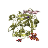
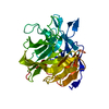
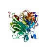
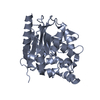
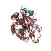
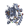
| Title | Crystal Structure of dimethylated RSL - sulfonatocalix[4]arene complex | |||||||||
 Components Components | Fucose-binding lectin protein | |||||||||
 Keywords Keywords | SUGAR BINDING PROTEIN /  calixarene / dimethyllysine / supramolecular recognition calixarene / dimethyllysine / supramolecular recognition | |||||||||
| Function / homology |  Function and homology information Function and homology information | |||||||||
| Biological species |  Ralstonia solanacearum (bacteria) Ralstonia solanacearum (bacteria) | |||||||||
| Method | X-RAY DIFFRACTION / MOLECULAR REPLACEMENT / Resolution: 1.6 Å | |||||||||
 Authors Authors | Guagnini, F. / Rennie, M.L. / Crowley, P.B. | |||||||||
| Funding support |  Ireland, 1items Ireland, 1items
| |||||||||
 Citation Citation | Journal: To Be Published Title: Protein Crystallization via Sulfonatocalix[4]arene Dimethylammonium Complexation Authors: Guagnini, F. / Antonik, P.M. / Rennie, M.L. / Pinalli, R. / Dalcanale, E. / Crowley, P.B. | |||||||||
| History |
|
- Structure visualization
| Structure viewer | Molecule: MolmilJmol/JSmol |
|---|
- Downloads & links
-Download
| PDBx/mmCIF format | 6gl5.cif.gz | 88.2 KB | Display | PDBx/mmCIF format |
|---|---|---|---|---|
| PDB format | pdb6gl5.ent.gz | 67.1 KB | Display | PDB format |
| PDBx/mmJSON format | 6gl5.json.gz | Tree view | PDBx/mmJSON format | |
| Others |  Other downloads Other downloads |
-Validation report
| Arichive directory | https://data.pdbj.org/pub/pdb/validation_reports/gl/6gl5ftp://data.pdbj.org/pub/pdb/validation_reports/gl/6gl5 | HTTPS FTP |
|---|
-Related structure data
| Related structure data | 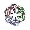 2bt9S S: Starting model for refinement |
|---|---|
| Similar structure data |


-Links
 PDBj
PDBj
- Assembly
| Deposited unit | 
| |||||||||||||||||||||||||||||||||||||||||||||||||||||
|---|---|---|---|---|---|---|---|---|---|---|---|---|---|---|---|---|---|---|---|---|---|---|---|---|---|---|---|---|---|---|---|---|---|---|---|---|---|---|---|---|---|---|---|---|---|---|---|---|---|---|---|---|---|---|
| 1 |
| |||||||||||||||||||||||||||||||||||||||||||||||||||||
| Unit cell |
| |||||||||||||||||||||||||||||||||||||||||||||||||||||
| Noncrystallographic symmetry (NCS) | NCS domain:
NCS domain segments: Component-ID: 0 / Beg auth comp-ID: SER / Beg label comp-ID: SER / End auth comp-ID: ASN / End label comp-ID: ASN / Refine code: 0 / Auth seq-ID: 2 - 90 / Label seq-ID: 2 - 90
NCS ensembles :
|
-Components
-Protein , 1 types, 3 molecules ABC
| #1: Protein | Mass: 9842.753 Da / Num. of mol.: 3 / Mutation: S88A Source method: isolated from a genetically manipulated source Details: Recombinant protein dimethylated at lysine residues and N-terminus Source: (gene. exp.) Ralstonia solanacearum (bacteria)Gene: E7Z57_08365, RSP795_21825, RSP822_19650, RUN39_v1_50103 Production host: Escherichia coli BL21(DE3) (bacteria) / References: UniProt: A0A0S4TLR1 |
|---|
-Non-polymers , 7 types, 394 molecules 


| #2: Chemical | ChemComp-T3Y /  Mass: 744.741 Da / Num. of mol.: 6 / Source method: obtained synthetically / Formula: C28H24O16S4 / Feature type: SUBJECT OF INVESTIGATION Mass: 744.741 Da / Num. of mol.: 6 / Source method: obtained synthetically / Formula: C28H24O16S4 / Feature type: SUBJECT OF INVESTIGATION#3: Chemical | ChemComp-GOL / Glycerol Mass: 92.094 Da / Num. of mol.: 6 / Source method: obtained synthetically / Formula: C3H8O3 Mass: 92.094 Da / Num. of mol.: 6 / Source method: obtained synthetically / Formula: C3H8O3#4: Chemical | ChemComp-PG4 / | Polyethylene glycol Mass: 194.226 Da / Num. of mol.: 1 / Source method: obtained synthetically / Formula: C8H18O5 / Comment: precipitant*YM Mass: 194.226 Da / Num. of mol.: 1 / Source method: obtained synthetically / Formula: C8H18O5 / Comment: precipitant*YM#5: Chemical | ChemComp-PEG / Diethylene glycol Mass: 106.120 Da / Num. of mol.: 4 / Source method: obtained synthetically / Formula: C4H10O3 Mass: 106.120 Da / Num. of mol.: 4 / Source method: obtained synthetically / Formula: C4H10O3#6: Chemical | ChemComp-EDO / | Ethylene glycol Mass: 62.068 Da / Num. of mol.: 1 / Source method: obtained synthetically / Formula: C2H6O2 Mass: 62.068 Da / Num. of mol.: 1 / Source method: obtained synthetically / Formula: C2H6O2#7: Chemical | ChemComp-SO4 / | Sulfate Mass: 96.063 Da / Num. of mol.: 1 / Source method: obtained synthetically / Formula: SO4 Mass: 96.063 Da / Num. of mol.: 1 / Source method: obtained synthetically / Formula: SO4#8: Water | ChemComp-HOH / | WaterMass: 18.015 Da / Num. of mol.: 375 / Source method: isolated from a natural source / Formula: H2O |
|---|
-Experimental details
-Experiment
| Experiment | Method: X-RAY DIFFRACTION / Number of used crystals: 1 |
|---|
- Sample preparation
| Crystal | Density Matthews: 2.71 Å3/Da / Density % sol: 54.56 % |
|---|---|
| Crystal grow | Temperature: 293 K / Method: vapor diffusion, sitting drop / pH: 4 / Details: 0.8 M ammonium sulfate, 0.1 M sodium citrate |
-Data collection
| Diffraction | Mean temperature: 100 K | |||||||||||||||||||||
|---|---|---|---|---|---|---|---|---|---|---|---|---|---|---|---|---|---|---|---|---|---|---|
| Diffraction source | Source: ROTATING ANODE / Type: RIGAKU / Wavelength: 1.5418 Å | |||||||||||||||||||||
| Detector | Type: RIGAKU RAXIS IV++ / Detector: IMAGE PLATE / Date: Apr 3, 2017 | |||||||||||||||||||||
| Radiation | Protocol: SINGLE WAVELENGTH / Monochromatic (M) / Laue (L): M / Scattering type: x-ray | |||||||||||||||||||||
| Radiation wavelength | Wavelength: 1.5418 Å / Relative weight: 1 | |||||||||||||||||||||
| Reflection | Resolution: 1.6→45.94 Å / Num. obs: 42850 / % possible obs: 99.5 % / Redundancy: 6.8 % / CC1/2: 0.999 / Rmerge(I) obs: 0.051 / Net I/σ(I): 24.1 | |||||||||||||||||||||
| Reflection shell |
|
- Processing
| Software |
| ||||||||||||||||||||||||||||||||||||||||||||||||||||||||||||
|---|---|---|---|---|---|---|---|---|---|---|---|---|---|---|---|---|---|---|---|---|---|---|---|---|---|---|---|---|---|---|---|---|---|---|---|---|---|---|---|---|---|---|---|---|---|---|---|---|---|---|---|---|---|---|---|---|---|---|---|---|---|
| Refinement | Method to determine structure: MOLECULAR REPLACEMENT Starting model: 2BT9 chain A Resolution: 1.6→38.02 Å / Cor.coef. Fo:Fc: 0.964 / Cor.coef. Fo:Fc free: 0.956 / SU B: 1.314 / SU ML: 0.046 / Cross valid method: THROUGHOUT / σ(F): 0 / ESU R: 0.084 / ESU R Free: 0.08 Details: HYDROGENS HAVE BEEN ADDED IN THE RIDING POSITIONS U VALUES : REFINED INDIVIDUALLY
| ||||||||||||||||||||||||||||||||||||||||||||||||||||||||||||
| Solvent computation | Ion probe radii: 0.8 Å / Shrinkage radii: 0.8 Å / VDW probe radii: 1.2 Å | ||||||||||||||||||||||||||||||||||||||||||||||||||||||||||||
| Displacement parameters | Biso max: 67.52 Å2 / Biso mean: 17.219 Å2 / Biso min: 7.66 Å2
| ||||||||||||||||||||||||||||||||||||||||||||||||||||||||||||
| Refinement step | Cycle: final / Resolution: 1.6→38.02 Å
| ||||||||||||||||||||||||||||||||||||||||||||||||||||||||||||
| Refine LS restraints |
| ||||||||||||||||||||||||||||||||||||||||||||||||||||||||||||
| Refine LS restraints NCS | Refine-ID: X-RAY DIFFRACTION / Type: interatomic distance / Weight position: 0.05
| ||||||||||||||||||||||||||||||||||||||||||||||||||||||||||||
| LS refinement shell | Resolution: 1.6→1.642 Å / Rfactor Rfree error: 0 / Total num. of bins used: 20
|
