 Movie
Movie Controller

[English] 日本語
 Yorodumi
- PDB-6gfg: Inositol 1,3,4,5,6-pentakisphosphate 2-kinase from A. thaliana in... -
+ Open data

- Basic information
| Entry | Database: PDB / ID: 6gfg | ||||||
|---|---|---|---|---|---|---|---|
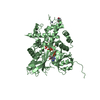
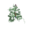
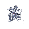
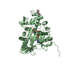
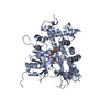
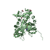
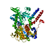
| Title | Inositol 1,3,4,5,6-pentakisphosphate 2-kinase from A. thaliana in complex with D-chiro-IP6 and ADP | ||||||
 Components Components | Inositol-pentakisphosphate 2-kinase | ||||||
 Keywords Keywords | TRANSFERASE / IPK1 / MYO-IP5 | ||||||
| Function / homology |  Function and homology information Function and homology informationinositol tetrakisphosphate 2-kinase activity / inositol-pentakisphosphate 2-kinase / inositol pentakisphosphate 2-kinase activity / myo-inositol hexakisphosphate biosynthetic process / lateral root development / intracellular phosphate ion homeostasis / phosphate ion homeostasis / defense response to fungus / defense response to virus / defense response to bacterium ...inositol tetrakisphosphate 2-kinase activity / inositol-pentakisphosphate 2-kinase / inositol pentakisphosphate 2-kinase activity / myo-inositol hexakisphosphate biosynthetic process / lateral root development / intracellular phosphate ion homeostasis / phosphate ion homeostasis / defense response to fungus / defense response to virus / defense response to bacterium / phosphorylation / ATP binding / metal ion binding / nucleus / cytoplasmSimilarity search - Function | ||||||
| Biological species |  Arabidopsis thaliana (thale cress) Arabidopsis thaliana (thale cress) | ||||||
| Method | X-RAY DIFFRACTION / SYNCHROTRON / MOLECULAR REPLACEMENT / Resolution: 3 Å | ||||||
 Authors Authors | Whitfield, H.L. / Brearley, C.A. / Hemmings, A.M. | ||||||
 Citation Citation | Journal: J. Med. Chem. / Year: 2018 Title: A Fluorescent Probe Identifies Active Site Ligands of Inositol Pentakisphosphate 2-Kinase. Authors: Whitfield, H. / Gilmartin, M. / Baker, K. / Riley, A.M. / Godage, H.Y. / Potter, B.V.L. / Hemmings, A.M. / Brearley, C.A. | ||||||
| History |
|
- Structure visualization
| Structure viewer | Molecule: MolmilJmol/JSmol |
|---|
- Downloads & links
-Download
| PDBx/mmCIF format | 6gfg.cif.gz | 187.5 KB | Display | PDBx/mmCIF format |
|---|---|---|---|---|
| PDB format | pdb6gfg.ent.gz | 145.4 KB | Display | PDB format |
| PDBx/mmJSON format | 6gfg.json.gz | Tree view | PDBx/mmJSON format | |
| Others |  Other downloads Other downloads |
-Validation report
| Arichive directory | https://data.pdbj.org/pub/pdb/validation_reports/gf/6gfgftp://data.pdbj.org/pub/pdb/validation_reports/gf/6gfg | HTTPS FTP |
|---|
-Related structure data
| Related structure data | 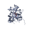 6fjkC 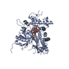 6fl3C  6fl8C  6gfhC  2xamS S: Starting model for refinement C: citing same article ( |
|---|---|
| Similar structure data |


-Links
 PDBj
PDBj- Assembly
| Deposited unit | 
| ||||||||
|---|---|---|---|---|---|---|---|---|---|
| 1 | 
| ||||||||
| 2 | 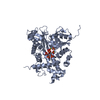
| ||||||||
| Unit cell |
|
-Components
| #1: Protein | Mass: 52863.969 Da / Num. of mol.: 2 Source method: isolated from a genetically manipulated source Source: (gene. exp.) Arabidopsis thaliana (thale cress) / Gene: AXX17_At5g40720 / Production host:  Escherichia coli (E. coli) Escherichia coli (E. coli)References: UniProt: A0A178UAB5, UniProt: Q93YN9*PLUS, inositol-pentakisphosphate 2-kinase#2: Chemical |   Mass: 660.035 Da / Num. of mol.: 2 / Source method: obtained synthetically / Formula: C6H18O24P6 Mass: 660.035 Da / Num. of mol.: 2 / Source method: obtained synthetically / Formula: C6H18O24P6#3: Chemical | Adenosine diphosphate  Mass: 427.201 Da / Num. of mol.: 2 / Source method: obtained synthetically / Formula: C10H15N5O10P2 / Comment: ADP, energy-carrying molecule*YM Mass: 427.201 Da / Num. of mol.: 2 / Source method: obtained synthetically / Formula: C10H15N5O10P2 / Comment: ADP, energy-carrying molecule*YM#4: Chemical | ChemComp-MG /   Mass: 24.305 Da / Num. of mol.: 4 / Source method: obtained synthetically / Formula: Mg Mass: 24.305 Da / Num. of mol.: 4 / Source method: obtained synthetically / Formula: Mg#5: Chemical |   Mass: 65.409 Da / Num. of mol.: 2 / Source method: obtained synthetically / Formula: Zn Mass: 65.409 Da / Num. of mol.: 2 / Source method: obtained synthetically / Formula: Zn |
|---|
-Experimental details
-Experiment
| Experiment | Method: X-RAY DIFFRACTION / Number of used crystals: 1 |
|---|
- Sample preparation
| Crystal | Density Matthews: 2.51 Å3/Da / Density % sol: 50.91 % |
|---|---|
| Crystal grow | Temperature: 289 K / Method: vapor diffusion, sitting drop / pH: 6.5 Details: 18 % PEG 3350, 0.1 M bis-tris propane pH 6.5, 2 mM MgCl2, 25% EG or 35% PEG 3350, 0.1 M bis-tris propane pH 6.5, 2 mM MgCl2 |
-Data collection
| Diffraction | Mean temperature: 100 K | ||||||||||||||||||||||||
|---|---|---|---|---|---|---|---|---|---|---|---|---|---|---|---|---|---|---|---|---|---|---|---|---|---|
| Diffraction source | Source: SYNCHROTRON / Site: Diamond  / Beamline: I04 / Wavelength: 0.9795 Å / Beamline: I04 / Wavelength: 0.9795 Å | ||||||||||||||||||||||||
| Detector | Type: ADSC QUANTUM 315 / Detector: CCD / Date: Feb 16, 2012 | ||||||||||||||||||||||||
| Radiation | Protocol: SINGLE WAVELENGTH / Monochromatic (M) / Laue (L): M / Scattering type: x-ray | ||||||||||||||||||||||||
| Radiation wavelength | Wavelength: 0.9795 Å / Relative weight: 1 | ||||||||||||||||||||||||
| Reflection | Resolution: 3→29.02 Å / Num. obs: 19896 / % possible obs: 96.7 % / Redundancy: 2.3 % / Biso Wilson estimate: 55.76 Å2 / CC1/2: 0.953 / Rmerge(I) obs: 0.149 / Rpim(I) all: 0.131 / Rrim(I) all: 0.199 / Net I/σ(I): 7.1 / Num. measured all: 45894 | ||||||||||||||||||||||||
| Reflection shell | Diffraction-ID: 1
|
- Processing
| Software |
| |||||||||||||||||||||||||||||||||||||||||||||||||||||||||||||||
|---|---|---|---|---|---|---|---|---|---|---|---|---|---|---|---|---|---|---|---|---|---|---|---|---|---|---|---|---|---|---|---|---|---|---|---|---|---|---|---|---|---|---|---|---|---|---|---|---|---|---|---|---|---|---|---|---|---|---|---|---|---|---|---|---|
| Refinement | Method to determine structure: MOLECULAR REPLACEMENT Starting model: 2XAM Resolution: 3→29.017 Å / SU ML: 0.5 / Cross valid method: THROUGHOUT / σ(F): 1.96 / Phase error: 28.87
| |||||||||||||||||||||||||||||||||||||||||||||||||||||||||||||||
| Solvent computation | Shrinkage radii: 0.9 Å / VDW probe radii: 1.11 Å | |||||||||||||||||||||||||||||||||||||||||||||||||||||||||||||||
| Displacement parameters | Biso max: 164.4 Å2 / Biso mean: 52.4431 Å2 / Biso min: 17.04 Å2 | |||||||||||||||||||||||||||||||||||||||||||||||||||||||||||||||
| Refinement step | Cycle: final / Resolution: 3→29.017 Å
| |||||||||||||||||||||||||||||||||||||||||||||||||||||||||||||||
| Refine LS restraints |
| |||||||||||||||||||||||||||||||||||||||||||||||||||||||||||||||
| LS refinement shell | Refine-ID: X-RAY DIFFRACTION / Rfactor Rfree error: 0 / Total num. of bins used: 8
|
