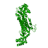
 Movie
Movie Controller


+ Open data

- Basic information
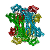
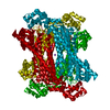
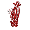
| Entry | Database: PDB / ID: 6g3g | ||||||
|---|---|---|---|---|---|---|---|
| Title | Crystal structure of EDDS lyase in complex with succinate | ||||||
 Components Components | Argininosuccinate lyase | ||||||
 Keywords Keywords | LYASE / C-N Lyase / metal chelator / EDDS / tetramer / succinate / aspartase fumarase superfamily | ||||||
| Function / homology |  Function and homology informationargininosuccinate lyase / argininosuccinate lyase activity / arginine biosynthetic process via ornithine Function and homology informationargininosuccinate lyase / argininosuccinate lyase activity / arginine biosynthetic process via ornithineSimilarity search - Function | ||||||
| Biological species | Chelativorans sp. | ||||||
| Method | X-RAY DIFFRACTION / MOLECULAR REPLACEMENT / Resolution: 2.606 Å | ||||||
 Authors Authors | Poddar, H. / Thunnissem, A.M.W.H. / Poelarends, G.J. | ||||||
| Funding support |  Netherlands, 1items Netherlands, 1items
| ||||||
 Citation Citation | Journal: Biochemistry / Year: 2018 Title: Structural Basis for the Catalytic Mechanism of Ethylenediamine- N, N'-disuccinic Acid Lyase, a Carbon-Nitrogen Bond-Forming Enzyme with a Broad Substrate Scope. Authors: Poddar, H. / de Villiers, J. / Zhang, J. / Puthan Veetil, V. / Raj, H. / Thunnissen, A.W.H. / Poelarends, G.J. | ||||||
| History |
|
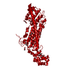
- Structure visualization
| Structure viewer | Molecule: MolmilJmol/JSmol |
|---|
- Downloads & links
-Download
| PDBx/mmCIF format | 6g3g.cif.gz | 110.7 KB | Display | PDBx/mmCIF format |
|---|---|---|---|---|
| PDB format | pdb6g3g.ent.gz | 84.7 KB | Display | PDB format |
| PDBx/mmJSON format | 6g3g.json.gz | Tree view | PDBx/mmJSON format | |
| Others |  Other downloads Other downloads |
-Validation report
| Arichive directory | https://data.pdbj.org/pub/pdb/validation_reports/g3/6g3gftp://data.pdbj.org/pub/pdb/validation_reports/g3/6g3g | HTTPS FTP |
|---|
-Related structure data
| Related structure data |  6g3dSC 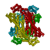 6g3eC  6g3fC  6g3hC  6g3iC S: Starting model for refinement C: citing same article ( |
|---|---|
| Similar structure data |
-Links
 PDBj
PDBj

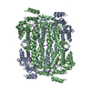
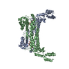
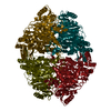
- Assembly
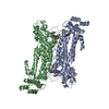
| Deposited unit | 
| ||||||||
|---|---|---|---|---|---|---|---|---|---|
| 1 | 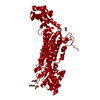
| ||||||||
| Unit cell |
| ||||||||
| Components on special symmetry positions |
|
-Components
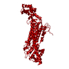
| #1: Protein | Mass: 55758.246 Da / Num. of mol.: 1 Source method: isolated from a genetically manipulated source Source: (gene. exp.)  Chelativorans sp. (strain BNC1) (bacteria) Chelativorans sp. (strain BNC1) (bacteria)Strain: BNC1 / Gene: Meso_0564 / Plasmid: pBADN / Production host: Escherichia coli (E. coli) / References: UniProt: Q11KV9 | ||
|---|---|---|---|
| #2: Chemical | ChemComp-SIN / Succinic acid  Mass: 118.088 Da / Num. of mol.: 1 / Source method: obtained synthetically / Formula: C4H6O4 Mass: 118.088 Da / Num. of mol.: 1 / Source method: obtained synthetically / Formula: C4H6O4 | ||
| #3: Chemical | ChemComp-PEG / Diethylene glycol  Mass: 106.120 Da / Num. of mol.: 8 / Source method: obtained synthetically / Formula: C4H10O3 Mass: 106.120 Da / Num. of mol.: 8 / Source method: obtained synthetically / Formula: C4H10O3#4: Water | ChemComp-HOH / | Water Mass: 18.015 Da / Num. of mol.: 114 / Source method: isolated from a natural source / Formula: H2O Mass: 18.015 Da / Num. of mol.: 114 / Source method: isolated from a natural source / Formula: H2O |
-Experimental details
-Experiment
| Experiment | Method: X-RAY DIFFRACTION / Number of used crystals: 1 |
|---|
- Sample preparation
| Crystal | Density Matthews: 3.59 Å3/Da / Density % sol: 65.7 % |
|---|---|
| Crystal grow | Temperature: 295 K / Method: vapor diffusion, sitting drop / pH: 6.5 Details: 0.1 M Sodium cacodylate pH 6.5, 0.2 - 0.3 M sodium succinate |
-Data collection
| Diffraction | Mean temperature: 110 K |
|---|---|
| Diffraction source | Source: ROTATING ANODE / Type: BRUKER AXS MICROSTAR / Wavelength: 1.54 Å |
| Detector | Type: MAR scanner 345 mm plate / Detector: IMAGE PLATE / Date: May 12, 2013 |
| Radiation | Protocol: SINGLE WAVELENGTH / Monochromatic (M) / Laue (L): M / Scattering type: x-ray |
| Radiation wavelength | Wavelength: 1.54 Å / Relative weight: 1 |
| Reflection | Resolution: 2.6→52.5 Å / Num. obs: 24456 / % possible obs: 99.5 % / Redundancy: 4.9 % / Rmerge(I) obs: 0.06 / Net I/σ(I): 28.5 |
| Reflection shell | Resolution: 2.6→2.71 Å |
- Processing
| Software |
| ||||||||||||||||||||||||||||||||||||||||||||||||||||||||||||||||||||||
|---|---|---|---|---|---|---|---|---|---|---|---|---|---|---|---|---|---|---|---|---|---|---|---|---|---|---|---|---|---|---|---|---|---|---|---|---|---|---|---|---|---|---|---|---|---|---|---|---|---|---|---|---|---|---|---|---|---|---|---|---|---|---|---|---|---|---|---|---|---|---|---|
| Refinement | Method to determine structure: MOLECULAR REPLACEMENT Starting model: 6G3D Resolution: 2.606→51.446 Å / SU ML: 0.28 / Cross valid method: FREE R-VALUE / σ(F): 1.35 / Phase error: 21.46
| ||||||||||||||||||||||||||||||||||||||||||||||||||||||||||||||||||||||
| Solvent computation | Shrinkage radii: 0.9 Å / VDW probe radii: 1.11 Å | ||||||||||||||||||||||||||||||||||||||||||||||||||||||||||||||||||||||
| Refinement step | Cycle: LAST / Resolution: 2.606→51.446 Å
| ||||||||||||||||||||||||||||||||||||||||||||||||||||||||||||||||||||||
| Refine LS restraints |
| ||||||||||||||||||||||||||||||||||||||||||||||||||||||||||||||||||||||
| LS refinement shell |
|
