 Movie
Movie Controller

[English] 日本語
 Yorodumi
- PDB-6fzw: Crystal structure of the metalloproteinase enhancer PCPE-1 bound ... -
+ Open data

- Basic information
| Entry | Database: PDB / ID: 6fzw | ||||||
|---|---|---|---|---|---|---|---|
| Title | Crystal structure of the metalloproteinase enhancer PCPE-1 bound to the procollagen C propeptide trimer (long) | ||||||
 Components Components |
| ||||||
 Keywords Keywords |  STRUCTURAL PROTEIN / Collagen STRUCTURAL PROTEIN / Collagen | ||||||
| Function / homology |  Function and homology information Function and homology informationcollagen type III trimer / aorta smooth muscle tissue morphogenesis / limb joint morphogenesis / transforming growth factor beta1 production / Crosslinking of collagen fibrils / collagen biosynthetic process / elastic fiber assembly / negative regulation of neuron migration / Collagen chain trimerization / platelet-derived growth factor binding ...collagen type III trimer / aorta smooth muscle tissue morphogenesis / limb joint morphogenesis / transforming growth factor beta1 production / Crosslinking of collagen fibrils / collagen biosynthetic process / elastic fiber assembly / negative regulation of neuron migration / Collagen chain trimerization / platelet-derived growth factor binding / endochondral bone morphogenesis / extracellular matrix structural constituent conferring tensile strength / basement membrane organization / Extracellular matrix organization / layer formation in cerebral cortex / Collagen biosynthesis and modifying enzymes / peptidase activator activity / peptide cross-linking / tissue homeostasis / Signaling by PDGF / negative regulation of immune response / NCAM1 interactions / digestive tract development / response to angiotensin / collagen fibril organization / Scavenging by Class A Receptors / extracellular matrix structural constituent / skin development / MET activates PTK2 signaling / Assembly of collagen fibrils and other multimeric structures / Syndecan interactions / positive regulation of Rho protein signal transduction / SMAD binding / Collagen degradation / Non-integrin membrane-ECM interactions / ECM proteoglycans / Integrin cell surface interactions / chondrocyte differentiation / supramolecular fiber organization / collagen binding / extracellular matrix organization / cell-matrix adhesion / transforming growth factor beta receptor signaling pathway / response to cytokine / integrin-mediated signaling pathway / cellular response to amino acid stimulus / neuron migration / lung development / wound healing / response to radiation / multicellular organism growth / cerebral cortex development / platelet activation / Immunoregulatory interactions between a Lymphoid and a non-Lymphoid cell / integrin binding / heparin binding / heart development / fibroblast proliferation / collagen-containing extracellular matrix / in utero embryonic development / protease binding / endoplasmic reticulum lumen / proteolysis / extracellular space / extracellular exosome / extracellular region / metal ion bindingSimilarity search - Function | ||||||
| Biological species |  Homo sapiens (human) Homo sapiens (human) | ||||||
| Method | X-RAY DIFFRACTION / SYNCHROTRON / MOLECULAR REPLACEMENT / Resolution: 2.78 Å | ||||||
 Authors Authors | Hohenester, E. / Pulido, D. | ||||||
| Funding support |  United Kingdom, 1items United Kingdom, 1items
| ||||||
 Citation Citation | Journal: Structure / Year: 2018 Title: Structural Basis for the Acceleration of Procollagen Processing by Procollagen C-Proteinase Enhancer-1. Authors: Pulido, D. / Sharma, U. / Vadon-Le Goff, S. / Hussain, S.A. / Cordes, S. / Mariano, N. / Bettler, E. / Moali, C. / Aghajari, N. / Hohenester, E. / Hulmes, D.J.S. | ||||||
| History |
|
- Structure visualization
| Structure viewer | Molecule: MolmilJmol/JSmol |
|---|
- Downloads & links
-Download
| PDBx/mmCIF format | 6fzw.cif.gz | 502 KB | Display | PDBx/mmCIF format |
|---|---|---|---|---|
| PDB format | pdb6fzw.ent.gz | 421 KB | Display | PDB format |
| PDBx/mmJSON format | 6fzw.json.gz | Tree view | PDBx/mmJSON format | |
| Others |  Other downloads Other downloads |
-Validation report
| Arichive directory | https://data.pdbj.org/pub/pdb/validation_reports/fz/6fzwftp://data.pdbj.org/pub/pdb/validation_reports/fz/6fzw | HTTPS FTP |
|---|
-Related structure data
| Related structure data | 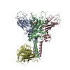 6fzvSC S: Starting model for refinement C: citing same article ( |
|---|---|
| Similar structure data |


-Links
 PDBj
PDBj


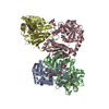
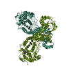
- Assembly
| Deposited unit | 
| ||||||||
|---|---|---|---|---|---|---|---|---|---|
| 1 |
| ||||||||
| Unit cell |
|
-Components
| #1: Protein | Mass: 31997.879 Da / Num. of mol.: 3 Source method: isolated from a genetically manipulated source Source: (gene. exp.) Homo sapiens (human) / Gene: COL3A1 / Plasmid: pHLsec / Production host: Homo sapiens (human) / References: UniProt: P02461#2: Protein | | Mass: 28696.900 Da / Num. of mol.: 1 Source method: isolated from a genetically manipulated source Source: (gene. exp.) Homo sapiens (human) / Gene: PCOLCE, PCPE1 / Plasmid: pCEP-Pu / Production host: Homo sapiens (human) / References: UniProt: Q15113#3: Chemical | ChemComp-CA /   Mass: 40.078 Da / Num. of mol.: 5 / Source method: obtained synthetically / Formula: Ca Mass: 40.078 Da / Num. of mol.: 5 / Source method: obtained synthetically / Formula: Ca#4: Chemical | Citric acid  Mass: 189.100 Da / Num. of mol.: 3 / Source method: obtained synthetically / Formula: C6H5O7 Mass: 189.100 Da / Num. of mol.: 3 / Source method: obtained synthetically / Formula: C6H5O7 |
|---|
-Experimental details
-Experiment
| Experiment | Method: X-RAY DIFFRACTION / Number of used crystals: 1 |
|---|
- Sample preparation
| Crystal | Density Matthews: 4.01 Å3/Da / Density % sol: 69.34 % |
|---|---|
| Crystal grow | Temperature: 295 K / Method: vapor diffusion, hanging drop Details: 20 mg/ml protein in 20 mM HEPES pH 7.5, 180 mM NaCl, 2.5 mM calcium chloride; 0.2 M ammonium citrate, 18% PEG3350 |
-Data collection
| Diffraction | Mean temperature: 100 K |
|---|---|
| Diffraction source | Source: SYNCHROTRON / Site: Diamond / Beamline: I04-1 / Wavelength: 0.9282 Å |
| Detector | Type: DECTRIS PILATUS 6M / Detector: PIXEL / Date: Sep 11, 2016 |
| Radiation | Protocol: SINGLE WAVELENGTH / Monochromatic (M) / Laue (L): M / Scattering type: x-ray |
| Radiation wavelength | Wavelength: 0.9282 Å / Relative weight: 1 |
| Reflection | Resolution: 2.78→68.8 Å / Num. obs: 46452 / % possible obs: 91.4 % / Redundancy: 4.8 % / CC1/2: 0.997 / Rmerge(I) obs: 0.066 / Net I/σ(I): 13.1 |
| Reflection shell | Resolution: 2.78→2.85 Å / Redundancy: 5.1 % / Rmerge(I) obs: 0.983 / Mean I/σ(I) obs: 1.3 / CC1/2: 0.634 / % possible all: 94.9 |
- Processing
| Software |
| |||||||||||||||||||||||||||||||||||||||||||||||||||||||||||||||||||||||||||||||||||||||||||||||||||||||||||||||||||||||
|---|---|---|---|---|---|---|---|---|---|---|---|---|---|---|---|---|---|---|---|---|---|---|---|---|---|---|---|---|---|---|---|---|---|---|---|---|---|---|---|---|---|---|---|---|---|---|---|---|---|---|---|---|---|---|---|---|---|---|---|---|---|---|---|---|---|---|---|---|---|---|---|---|---|---|---|---|---|---|---|---|---|---|---|---|---|---|---|---|---|---|---|---|---|---|---|---|---|---|---|---|---|---|---|---|---|---|---|---|---|---|---|---|---|---|---|---|---|---|---|---|
| Refinement | Method to determine structure: MOLECULAR REPLACEMENT Starting model: 6FZV Resolution: 2.78→68.787 Å / SU ML: 0.48 / Cross valid method: FREE R-VALUE / σ(F): 1.34 / Phase error: 32.3
| |||||||||||||||||||||||||||||||||||||||||||||||||||||||||||||||||||||||||||||||||||||||||||||||||||||||||||||||||||||||
| Solvent computation | Shrinkage radii: 0.9 Å / VDW probe radii: 1.11 Å | |||||||||||||||||||||||||||||||||||||||||||||||||||||||||||||||||||||||||||||||||||||||||||||||||||||||||||||||||||||||
| Refinement step | Cycle: LAST / Resolution: 2.78→68.787 Å
| |||||||||||||||||||||||||||||||||||||||||||||||||||||||||||||||||||||||||||||||||||||||||||||||||||||||||||||||||||||||
| Refine LS restraints |
| |||||||||||||||||||||||||||||||||||||||||||||||||||||||||||||||||||||||||||||||||||||||||||||||||||||||||||||||||||||||
| LS refinement shell |
| |||||||||||||||||||||||||||||||||||||||||||||||||||||||||||||||||||||||||||||||||||||||||||||||||||||||||||||||||||||||
| Refinement TLS params. | Method: refined / Origin x: 6.9696 Å / Origin y: -21.6768 Å / Origin z: 4.1677 Å
| |||||||||||||||||||||||||||||||||||||||||||||||||||||||||||||||||||||||||||||||||||||||||||||||||||||||||||||||||||||||
| Refinement TLS group | Selection details: all |
