 Movie
Movie Controller

[English] 日本語
 Yorodumi
- PDB-6fu2: ATP phosphoribosyltransferase (HisZG ATPPRT) from Psychrobacter a... -
+ Open data

- Basic information
| Entry | Database: PDB / ID: 6fu2 | ||||||
|---|---|---|---|---|---|---|---|
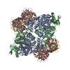
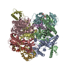
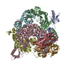
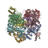
| Title | ATP phosphoribosyltransferase (HisZG ATPPRT) from Psychrobacter arcticus in complex with PRPP and ATP | ||||||
 Components Components |
| ||||||
 Keywords Keywords |  TRANSFERASE / ATP phosphoribosyltransferase / HisZG / cold-adapted TRANSFERASE / ATP phosphoribosyltransferase / HisZG / cold-adapted | ||||||
| Function / homology |  Function and homology informationATP phosphoribosyltransferase / ATP phosphoribosyltransferase activity / histidine biosynthetic process / ATP binding / cytoplasm Function and homology informationATP phosphoribosyltransferase / ATP phosphoribosyltransferase activity / histidine biosynthetic process / ATP binding / cytoplasmSimilarity search - Function | ||||||
| Biological species |  Psychrobacter arcticus (bacteria) Psychrobacter arcticus (bacteria) | ||||||
| Method | X-RAY DIFFRACTION / SYNCHROTRON / MOLECULAR REPLACEMENT / molecular replacement / Resolution: 2.71 Å | ||||||
 Authors Authors | Alphey, M.S. / Ge, Y. / Fisher, G. / Czekster, C.M. / Naismith, J.H. / da Silva, R.G. | ||||||
| Funding support |  United Kingdom, 1items United Kingdom, 1items
| ||||||
 Citation Citation | Journal: Acs Catalysis / Year: 2018 Title: Catalytic and Anticatalytic Snapshots of a Short-Form ATP Phosphoribosyltransferase Authors: Alphey, M.S. / Fisher, G. / Hirschi, J.S. / Stroek, R. / Ge, Y. / Gould, E.R. / Czekster, C.M. / Liu, H. / Florence, G.J. / Vetticatt, M.J. / Naismith, J.H. / da Silva, R.G. | ||||||
| History |
|
- Structure visualization
| Structure viewer | Molecule: MolmilJmol/JSmol |
|---|
- Downloads & links
-Download
| PDBx/mmCIF format | 6fu2.cif.gz | 241.3 KB | Display | PDBx/mmCIF format |
|---|---|---|---|---|
| PDB format | pdb6fu2.ent.gz | 190.1 KB | Display | PDB format |
| PDBx/mmJSON format | 6fu2.json.gz | Tree view | PDBx/mmJSON format | |
| Others |  Other downloads Other downloads |
-Validation report
| Arichive directory | https://data.pdbj.org/pub/pdb/validation_reports/fu/6fu2ftp://data.pdbj.org/pub/pdb/validation_reports/fu/6fu2 | HTTPS FTP |
|---|
-Related structure data
| Related structure data |  6fcaC  6fccC  6fctC  6fcwC  6fcyC  6fd9C  6fttC  6fu7C  6fuaC  5m8hS S: Starting model for refinement C: citing same article ( |
|---|---|
| Similar structure data |


-Links
 PDBj
PDBj

- Assembly
| Deposited unit | 
| ||||||||||||||||||||||||||||||||||||||||||||||||||||||||||||||||||||
|---|---|---|---|---|---|---|---|---|---|---|---|---|---|---|---|---|---|---|---|---|---|---|---|---|---|---|---|---|---|---|---|---|---|---|---|---|---|---|---|---|---|---|---|---|---|---|---|---|---|---|---|---|---|---|---|---|---|---|---|---|---|---|---|---|---|---|---|---|---|
| 1 | 
| ||||||||||||||||||||||||||||||||||||||||||||||||||||||||||||||||||||
| Unit cell |
| ||||||||||||||||||||||||||||||||||||||||||||||||||||||||||||||||||||
| Noncrystallographic symmetry (NCS) | NCS domain:
NCS domain segments: Component-ID: 0 / Refine code: 0
NCS ensembles :
|
-Components
-Protein , 2 types, 4 molecules ABCD
| #1: Protein | Mass: 43129.039 Da / Num. of mol.: 2 Source method: isolated from a genetically manipulated source Details: Missing sections are due to flexible regions not visible in the electron density maps. N-terminal Gly remains after tag cleavage. Source: (gene. exp.) Psychrobacter arcticus (bacteria) / Gene: hisZ, Psyc_0676 / Plasmid: pJexpress414 / Production host: Escherichia coli BL21(DE3) (bacteria) / References: UniProt: Q4FTX3#2: Protein | / ATP-PRTaseMass: 25240.965 Da / Num. of mol.: 2 Source method: isolated from a genetically manipulated source Details: Missing sections are due to flexible regions not visible in the electron density maps. N-terminal Gly remains after tag cleavage. Source: (gene. exp.) Psychrobacter arcticus (bacteria) / Gene: hisG, Psyc_1903 / Plasmid: pJexpress431 / Production host: Escherichia coli BL21(DE3) (bacteria) / Variant (production host): C43 / References: UniProt: Q4FQF7, ATP phosphoribosyltransferase |
|---|
-Sugars , 1 types, 2 molecules 
| #4: Sugar | Phosphoribosyl pyrophosphate Type: D-saccharide / Mass: 390.070 Da / Num. of mol.: 2 / Source method: obtained synthetically / Formula: C5H13O14P3 Type: D-saccharide / Mass: 390.070 Da / Num. of mol.: 2 / Source method: obtained synthetically / Formula: C5H13O14P3 |
|---|
-Non-polymers , 4 types, 16 molecules 


| #3: Chemical | Tris Mass: 122.143 Da / Num. of mol.: 3 / Source method: obtained synthetically / Formula: C4H12NO3 / Comment: pH buffer*YM Mass: 122.143 Da / Num. of mol.: 3 / Source method: obtained synthetically / Formula: C4H12NO3 / Comment: pH buffer*YM#5: Chemical | Adenosine triphosphate Mass: 507.181 Da / Num. of mol.: 2 / Source method: obtained synthetically / Formula: C10H16N5O13P3 / Comment: ATP, energy-carrying molecule*YM Mass: 507.181 Da / Num. of mol.: 2 / Source method: obtained synthetically / Formula: C10H16N5O13P3 / Comment: ATP, energy-carrying molecule*YM#6: Chemical |  Mass: 24.305 Da / Num. of mol.: 2 / Source method: obtained synthetically / Formula: Mg Mass: 24.305 Da / Num. of mol.: 2 / Source method: obtained synthetically / Formula: Mg#7: Water | ChemComp-HOH / | WaterMass: 18.015 Da / Num. of mol.: 9 / Source method: isolated from a natural source / Formula: H2O |
|---|
-Experimental details
-Experiment
| Experiment | Method: X-RAY DIFFRACTION / Number of used crystals: 1 |
|---|
- Sample preparation
| Crystal | Density Matthews: 2.54 Å3/Da / Density % sol: 51.51 % |
|---|---|
| Crystal grow | Temperature: 277 K / Method: vapor diffusion, hanging drop / pH: 8.5 Details: 8mg mL-1 protein (in 20mM Tris pH7, 50mM KCl, 10mM MgCl2, 2mM DTT, 0.5mM histidine) mixed in 1:1 ratio with precipitant solution (11% PEG3350, 100mM bicine pH8.5, 150mM SrCl2, 150mM KBr, 2% 1,6-hexanediol) |
-Data collection
| Diffraction | Mean temperature: 100 K |
|---|---|
| Diffraction source | Source: SYNCHROTRON / Site: Diamond / Beamline: I24 / Wavelength: 0.9686 Å |
| Detector | Type: DECTRIS PILATUS3 6M / Detector: PIXEL / Date: Apr 27, 2017 |
| Radiation | Protocol: SINGLE WAVELENGTH / Monochromatic (M) / Laue (L): M / Scattering type: x-ray |
| Radiation wavelength | Wavelength: 0.9686 Å / Relative weight: 1 |
| Reflection | Resolution: 2.71→73.11 Å / Num. obs: 36859 / % possible obs: 99.6 % / Redundancy: 4.1 % / Biso Wilson estimate: 64.8 Å2 / CC1/2: 0.996 / Rmerge(I) obs: 0.093 / Rpim(I) all: 0.052 / Rrim(I) all: 0.107 / Net I/σ(I): 10.8 |
| Reflection shell | Resolution: 2.71→2.78 Å / Redundancy: 4.2 % / Rmerge(I) obs: 1.095 / Num. unique obs: 2720 / CC1/2: 0.581 / Rpim(I) all: 0.59 / Rrim(I) all: 1.248 / % possible all: 99.7 |
-Phasing
| Phasing | Method: molecular replacement |
|---|
- Processing
| Software |
| ||||||||||||||||||||||||||||||||||||||||||||||||||||||||||||
|---|---|---|---|---|---|---|---|---|---|---|---|---|---|---|---|---|---|---|---|---|---|---|---|---|---|---|---|---|---|---|---|---|---|---|---|---|---|---|---|---|---|---|---|---|---|---|---|---|---|---|---|---|---|---|---|---|---|---|---|---|---|
| Refinement | Method to determine structure: MOLECULAR REPLACEMENT Starting model: 5M8H Resolution: 2.71→39.05 Å / Cor.coef. Fo:Fc: 0.956 / Cor.coef. Fo:Fc free: 0.932 / SU B: 16.129 / SU ML: 0.302 / Cross valid method: THROUGHOUT / σ(F): 0 / ESU R Free: 0.344 Details: HYDROGENS HAVE BEEN ADDED IN THE RIDING POSITIONS U VALUES : REFINED INDIVIDUALLY
| ||||||||||||||||||||||||||||||||||||||||||||||||||||||||||||
| Solvent computation | Ion probe radii: 0.7 Å / Shrinkage radii: 0.7 Å / VDW probe radii: 1 Å | ||||||||||||||||||||||||||||||||||||||||||||||||||||||||||||
| Displacement parameters | Biso max: 171.71 Å2 / Biso mean: 80.589 Å2 / Biso min: 47.55 Å2
| ||||||||||||||||||||||||||||||||||||||||||||||||||||||||||||
| Refinement step | Cycle: final / Resolution: 2.71→39.05 Å
| ||||||||||||||||||||||||||||||||||||||||||||||||||||||||||||
| Refine LS restraints |
| ||||||||||||||||||||||||||||||||||||||||||||||||||||||||||||
| Refine LS restraints NCS | Refine-ID: X-RAY DIFFRACTION / Type: interatomic distance / Rms dev position: 0.09 Å / Weight position: 0.05
| ||||||||||||||||||||||||||||||||||||||||||||||||||||||||||||
| LS refinement shell | Resolution: 2.71→2.78 Å / Rfactor Rfree error: 0 / Total num. of bins used: 20
|
