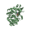
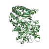
[English] 日本語
Yorodumi
- PDB-6fsk: F194Y mutant of the Dye-decolorizing peroxidase (DYP) from Pleuro... -

+
Open data


ID or keywords:

Loading...

-
Basic information

Entry
Database: PDB / ID: 6fsk
TitleF194Y mutant of the Dye-decolorizing peroxidase (DYP) from Pleurotus ostreatus
ComponentsDyP-type peroxidaseDye decolorizing peroxidase
KeywordsOXIDOREDUCTASE
Function / homologyDyP-type peroxidase family. / Dyp-type peroxidase / Dimeric alpha-beta barrel / peroxidase activity / heme binding / PROTOPORPHYRIN IX CONTAINING FE / DyP-type peroxidase
Function and homology information
Biological speciesPleurotus ostreatus PC15 (fungus)
MethodX-RAY DIFFRACTION / SYNCHROTRON / MOLECULAR REPLACEMENT / Resolution: 1.56 Å
AuthorsRomero, A. / Davo-Siguero, I.
Funding support Spain, 1items
OrganizationGrant numberCountry
Spanish Ministry of Economy and CompetitivenessBFU2016-77835-R Spain
CitationJournal: Acs Catalysis / Year: 2018
Title: Description of a non-canonical Mn(II)-oxidation site in peroxidases
Authors: Fernandez-Fueyo, E. / Davo-Siguero, I. / Almendral, D. / Linde, D. / Baratto, M.C. / Pogni, R. / Romero, A. / Guallar, V. / Martinez, A.T.
History
DepositionFeb 19, 2018Deposition site: PDBE / Processing site: PDBE
Revision 1.0Dec 26, 2018Provider: repository / Type: Initial release
Revision 2.0Jan 23, 2019Group: Advisory / Atomic model ...Advisory / Atomic model / Data collection / Derived calculations
Category: atom_site / pdbx_data_processing_status ...atom_site / pdbx_data_processing_status / pdbx_nonpoly_scheme / pdbx_solvent_atom_site_mapping / pdbx_validate_close_contact / struct_conn / struct_site / struct_site_gen
Item: _atom_site.B_iso_or_equiv / _atom_site.Cartn_x ..._atom_site.B_iso_or_equiv / _atom_site.Cartn_x / _atom_site.Cartn_y / _atom_site.Cartn_z / _atom_site.pdbx_auth_seq_id / _pdbx_nonpoly_scheme.auth_seq_num / _pdbx_solvent_atom_site_mapping.auth_seq_id
Revision 2.1Jan 17, 2024Group: Advisory / Data collection ...Advisory / Data collection / Database references / Derived calculations / Refinement description
Category: chem_comp_atom / chem_comp_bond ...chem_comp_atom / chem_comp_bond / database_2 / pdbx_initial_refinement_model / pdbx_unobs_or_zero_occ_residues / struct_conn
Item: _database_2.pdbx_DOI / _database_2.pdbx_database_accession ..._database_2.pdbx_DOI / _database_2.pdbx_database_accession / _struct_conn.conn_type_id / _struct_conn.id / _struct_conn.pdbx_dist_value / _struct_conn.pdbx_leaving_atom_flag / _struct_conn.ptnr1_auth_asym_id / _struct_conn.ptnr1_auth_comp_id / _struct_conn.ptnr1_auth_seq_id / _struct_conn.ptnr1_label_asym_id / _struct_conn.ptnr1_label_atom_id / _struct_conn.ptnr1_label_comp_id / _struct_conn.ptnr1_label_seq_id / _struct_conn.ptnr2_auth_asym_id / _struct_conn.ptnr2_auth_comp_id / _struct_conn.ptnr2_auth_seq_id / _struct_conn.ptnr2_label_asym_id / _struct_conn.ptnr2_label_atom_id / _struct_conn.ptnr2_label_comp_id / _struct_conn.ptnr2_label_seq_id

-
Structure visualization

Structure viewerMolecule:
MolmilJmol/JSmol

Downloads & links

-
Assembly

Deposited unit
A: DyP-type peroxidase
B: DyP-type peroxidase
hetero molecules


Theoretical massNumber of molelcules
Total (without water)107,7055
Polymers106,2772
Non-polymers1,4283
Water20,1591119
1
A: DyP-type peroxidase
hetero molecules


Theoretical massNumber of molelcules
Total (without water)53,9503
Polymers53,1391
Non-polymers8122
Water181
TypeNameSymmetry operationNumber
identity operation1_555x,y,z1
2
B: DyP-type peroxidase
hetero molecules


Theoretical massNumber of molelcules
Total (without water)53,7552
Polymers53,1391
Non-polymers6161
Water181
TypeNameSymmetry operationNumber
identity operation1_555x,y,z1
Unit cell
Length a, b, c (Å)62.717, 117.575, 140.434
Angle α, β, γ (deg.)90.00, 90.00, 90.00
Int Tables number19
Space group name H-MP212121

-
Components

#1: Protein DyP-type peroxidase / Dye decolorizing peroxidase


Mass: 53138.641 Da / Num. of mol.: 2 / Mutation: F194Y
Source method: isolated from a genetically manipulated source
Details: Cysteine at position 147 is oxidized to S-hydroxycysteine
Source: (gene. exp.) Pleurotus ostreatus PC15 (fungus) / Gene: DyP4, PLEOSDRAFT_1069077 / Production host: Escherichia coli #1/H766 (bacteria) / References: UniProt: A0A067N4E7
#2: Chemical ChemComp-HEM / PROTOPORPHYRIN IX CONTAINING FE / HEME / Heme B


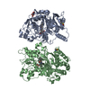
Mass: 616.487 Da / Num. of mol.: 2 / Source method: obtained synthetically / Formula: C34H32FeN4O4
#3: Chemical ChemComp-MES / 2-(N-MORPHOLINO)-ETHANESULFONIC ACID / MES (buffer)


Mass: 195.237 Da / Num. of mol.: 1 / Source method: obtained synthetically / Formula: C6H13NO4S / Comment: pH buffer*YM
#4: Water ChemComp-HOH / water / Water


Mass: 18.015 Da / Num. of mol.: 1119 / Source method: isolated from a natural source / Formula: H2O

-
Experimental details

-
Experiment

ExperimentMethod: X-RAY DIFFRACTION / Number of used crystals: 1

-
Sample preparation

CrystalDensity Matthews: 2.36 Å3/Da / Density % sol: 47.87 %
Crystal growTemperature: 295 K / Method: vapor diffusion, sitting drop / pH: 5.75 / Details: 0.2M MES pH=5.75 25%PEG 2000 MME

-
Data collection

DiffractionMean temperature: 110 K
Diffraction sourceSource: SYNCHROTRON / Site: ESRF / Beamline: ID30B / Wavelength: 0.976252 Å
DetectorType: DECTRIS PILATUS 6M / Detector: PIXEL / Date: Dec 12, 2015
RadiationMonochromator: Standard ESRF channel cut / Protocol: SINGLE WAVELENGTH / Monochromatic (M) / Laue (L): M / Scattering type: x-ray
Radiation wavelengthWavelength: 0.976252 Å / Relative weight: 1
ReflectionResolution: 1.56→70.22 Å / Num. obs: 144926 / % possible obs: 97.3 % / Observed criterion σ(F): 0 / Observed criterion σ(I): 0 / Redundancy: 4.5 % / Biso Wilson estimate: 17.7 Å2 / CC1/2: 0.997 / Rmerge(I) obs: 0.065 / Rpim(I) all: 0.051 / Rrim(I) all: 0.083 / Net I/σ(I): 12.7
Reflection shellResolution: 1.56→1.64 Å / Redundancy: 4.6 % / Rmerge(I) obs: 0.514 / Mean I/σ(I) obs: 2.3 / Num. unique obs: 20996 / CC1/2: 0.745 / Rpim(I) all: 0.392 / Rrim(I) all: 0.649 / % possible all: 97.4

-
Processing

Software
NameVersionClassification
REFMAC5.8.0155refinement
XDSdata reduction
Aimlessdata scaling
MOLREPphasing
RefinementMethod to determine structure: MOLECULAR REPLACEMENT
Starting model: 3AFV
Resolution: 1.56→70.22 Å / Cor.coef. Fo:Fc: 0.967 / Cor.coef. Fo:Fc free: 0.955 / SU B: 1.493 / SU ML: 0.053 / Cross valid method: THROUGHOUT / ESU R: 0.075 / ESU R Free: 0.08 / Details: HYDROGENS HAVE BEEN ADDED IN THE RIDING POSITIONS
RfactorNum. reflection% reflectionSelection details
Rfree0.19885 7250 5 %RANDOM
Rwork0.16201 ---
obs0.16386 137552 97.05 %-
Solvent computationIon probe radii: 0.8 Å / Shrinkage radii: 0.8 Å / VDW probe radii: 1.2 Å
Displacement parametersBiso mean: 18.56 Å2
Baniso -1Baniso -2Baniso -3
1-0.21 Å20 Å20 Å2
2--0.04 Å2-0 Å2
3----0.25 Å2
Refinement stepCycle: 1 / Resolution: 1.56→70.22 Å
ProteinNucleic acidLigandSolventTotal
Num. atoms7477 0 98 1119 8694
Refine LS restraints
Refine-IDTypeDev idealDev ideal targetNumber
X-RAY DIFFRACTIONr_bond_refined_d0.0210.0197733
X-RAY DIFFRACTIONr_bond_other_d0.0020.027232
X-RAY DIFFRACTIONr_angle_refined_deg2.0411.96610516
X-RAY DIFFRACTIONr_angle_other_deg1.237316681
X-RAY DIFFRACTIONr_dihedral_angle_1_deg6.1845955
X-RAY DIFFRACTIONr_dihedral_angle_2_deg34.03524.038364
X-RAY DIFFRACTIONr_dihedral_angle_3_deg11.166151197
X-RAY DIFFRACTIONr_dihedral_angle_4_deg23.2371548
X-RAY DIFFRACTIONr_chiral_restr0.1330.21105
X-RAY DIFFRACTIONr_gen_planes_refined0.0130.0218883
X-RAY DIFFRACTIONr_gen_planes_other0.0060.021827
X-RAY DIFFRACTIONr_nbd_refined
X-RAY DIFFRACTIONr_nbd_other
X-RAY DIFFRACTIONr_nbtor_refined
X-RAY DIFFRACTIONr_nbtor_other
X-RAY DIFFRACTIONr_xyhbond_nbd_refined
X-RAY DIFFRACTIONr_xyhbond_nbd_other
X-RAY DIFFRACTIONr_metal_ion_refined
X-RAY DIFFRACTIONr_metal_ion_other
X-RAY DIFFRACTIONr_symmetry_vdw_refined
X-RAY DIFFRACTIONr_symmetry_vdw_other
X-RAY DIFFRACTIONr_symmetry_hbond_refined
X-RAY DIFFRACTIONr_symmetry_hbond_other
X-RAY DIFFRACTIONr_symmetry_metal_ion_refined
X-RAY DIFFRACTIONr_symmetry_metal_ion_other
X-RAY DIFFRACTIONr_mcbond_it1.6641.6183829
X-RAY DIFFRACTIONr_mcbond_other1.6641.6183828
X-RAY DIFFRACTIONr_mcangle_it2.4122.4244781
X-RAY DIFFRACTIONr_mcangle_other2.4122.4244782
X-RAY DIFFRACTIONr_scbond_it2.761.8423904
X-RAY DIFFRACTIONr_scbond_other2.761.8423904
X-RAY DIFFRACTIONr_scangle_it
X-RAY DIFFRACTIONr_scangle_other4.1662.6575736
X-RAY DIFFRACTIONr_long_range_B_refined5.48820.8848995
X-RAY DIFFRACTIONr_long_range_B_other5.48820.8868996
X-RAY DIFFRACTIONr_rigid_bond_restr
X-RAY DIFFRACTIONr_sphericity_free
X-RAY DIFFRACTIONr_sphericity_bonded
LS refinement shellResolution: 1.556→1.597 Å / Total num. of bins used: 20
RfactorNum. reflection% reflection
Rfree0.334 553 -
Rwork0.307 10100 -
obs--97.39 %

+
About Yorodumi

-
News

-
Feb 9, 2022. New format data for meta-information of EMDB entries

New format data for meta-information of EMDB entries

  • Version 3 of the EMDB header file is now the official format.
  • The previous official version 1.9 will be removed from the archive.

Related info.:EMDB header

External links:wwPDB to switch to version 3 of the EMDB data model

-
Aug 12, 2020. Covid-19 info

Covid-19 info

URL: https://pdbj.org/emnavi/covid19.php

New page: Covid-19 featured information page in EM Navigator.

Related info.:Covid-19 info / Mar 5, 2020. Novel coronavirus structure data

+
Mar 5, 2020. Novel coronavirus structure data

Novel coronavirus structure data

Related info.:Yorodumi Speices / Aug 12, 2020. Covid-19 info

External links:COVID-19 featured content - PDBj / Molecule of the Month (242):Coronavirus Proteases

+
Jan 31, 2019. EMDB accession codes are about to change! (news from PDBe EMDB page)

EMDB accession codes are about to change! (news from PDBe EMDB page)

  • The allocation of 4 digits for EMDB accession codes will soon come to an end. Whilst these codes will remain in use, new EMDB accession codes will include an additional digit and will expand incrementally as the available range of codes is exhausted. The current 4-digit format prefixed with “EMD-” (i.e. EMD-XXXX) will advance to a 5-digit format (i.e. EMD-XXXXX), and so on. It is currently estimated that the 4-digit codes will be depleted around Spring 2019, at which point the 5-digit format will come into force.
  • The EM Navigator/Yorodumi systems omit the EMD- prefix.

Related info.:Q: What is EMD? / ID/Accession-code notation in Yorodumi/EM Navigator

External links:EMDB Accession Codes are Changing Soon! / Contact to PDBj

+
Jul 12, 2017. Major update of PDB

Major update of PDB

  • wwPDB released updated PDB data conforming to the new PDBx/mmCIF dictionary.
  • This is a major update changing the version number from 4 to 5, and with Remediation, in which all the entries are updated.
  • In this update, many items about electron microscopy experimental information are reorganized (e.g. em_software).
  • Now, EM Navigator and Yorodumi are based on the updated data.

External links:wwPDB Remediation / Enriched Model Files Conforming to OneDep Data Standards Now Available in the PDB FTP Archive

-
Yorodumi

Thousand views of thousand structures

  • Yorodumi is a browser for structure data from EMDB, PDB, SASBDB, etc.
  • This page is also the successor to EM Navigator detail page, and also detail information page/front-end page for Omokage search.
  • The word "yorodu" (or yorozu) is an old Japanese word meaning "ten thousand". "mi" (miru) is to see.

Related info.:EMDB / PDB / SASBDB / Comparison of 3 databanks / Yorodumi Search / Aug 31, 2016. New EM Navigator & Yorodumi / Yorodumi Papers / Jmol/JSmol / Function and homology information / Changes in new EM Navigator and Yorodumi

Read more