 Movie
Movie Controller

[English] 日本語
 Yorodumi
- PDB-6fsf: Crystal structure of the tandem PX-PH-domains of Bem3 from Saccha... -
+ Open data

- Basic information
| Entry | Database: PDB / ID: 6fsf | |||||||||
|---|---|---|---|---|---|---|---|---|---|---|
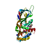
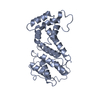
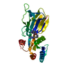
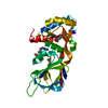
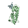
| Title | Crystal structure of the tandem PX-PH-domains of Bem3 from Saccharomyces cerevisiae | |||||||||
 Components Components | GTPase-activating protein BEM3 | |||||||||
 Keywords Keywords |  ENDOCYTOSIS / Bem3 / PX domain / PH domain / phox / pleckstrin homology / phosphatidylinositol phosphates / PIP ENDOCYTOSIS / Bem3 / PX domain / PH domain / phox / pleckstrin homology / phosphatidylinositol phosphates / PIP | |||||||||
| Function / homology |  Function and homology information Function and homology information: / : / RHOF GTPase cycle / CDC42 GTPase cycle / RHOD GTPase cycle / RHOV GTPase cycle / septin ring organization / RHOA GTPase cycle / incipient cellular bud site / cellular bud tip ...: / : / RHOF GTPase cycle / CDC42 GTPase cycle / RHOD GTPase cycle / RHOV GTPase cycle / septin ring organization / RHOA GTPase cycle / incipient cellular bud site / cellular bud tip / mating projection tip / phosphatidylinositol-3-phosphate binding / negative regulation of Rho protein signal transduction / establishment of cell polarity / regulation of GTPase activity / Neutrophil degranulation / GTPase activator activity / positive regulation of GTPase activity / cell cortex / signal transduction / cytoplasmSimilarity search - Function | |||||||||
| Biological species |  Saccharomyces cerevisiae S288C (yeast) Saccharomyces cerevisiae S288C (yeast) | |||||||||
| Method | X-RAY DIFFRACTION / SYNCHROTRON / SAD / Resolution: 2.2 Å | |||||||||
 Authors Authors | Ali, I. / Eu, S. / Koch, D. / Bleimling, N. / Goody, R.S. / Mueller, M.P. | |||||||||
| Funding support |  Germany, 2items Germany, 2items
| |||||||||
 Citation Citation | Journal: Acta Crystallogr F Struct Biol Commun / Year: 2018 Title: Structure of the tandem PX-PH domains of Bem3 from Saccharomyces cerevisiae. Authors: Ali, I. / Eu, S. / Koch, D. / Bleimling, N. / Goody, R.S. / Muller, M.P. | |||||||||
| History |
|
- Structure visualization
| Structure viewer | Molecule: MolmilJmol/JSmol |
|---|
- Downloads & links
-Download
| PDBx/mmCIF format | 6fsf.cif.gz | 104.6 KB | Display | PDBx/mmCIF format |
|---|---|---|---|---|
| PDB format | pdb6fsf.ent.gz | 80 KB | Display | PDB format |
| PDBx/mmJSON format | 6fsf.json.gz | Tree view | PDBx/mmJSON format | |
| Others |  Other downloads Other downloads |
-Validation report
| Arichive directory | https://data.pdbj.org/pub/pdb/validation_reports/fs/6fsfftp://data.pdbj.org/pub/pdb/validation_reports/fs/6fsf | HTTPS FTP |
|---|
-Related structure data
| Similar structure data |
|---|


-Links
 PDBj
PDBj


- Assembly
| Deposited unit | 
| ||||||||
|---|---|---|---|---|---|---|---|---|---|
| 1 |
| ||||||||
| Unit cell |
|
-Components
| #1: Protein | Mass: 30739.938 Da / Num. of mol.: 1 Source method: isolated from a genetically manipulated source Source: (gene. exp.) Saccharomyces cerevisiae S288C (yeast) / Gene: BEM3, YPL115C, LPH12C / Production host:  Escherichia coli (E. coli) / References: UniProt: P32873 Escherichia coli (E. coli) / References: UniProt: P32873 |
|---|---|
| #2: Water | ChemComp-HOH / Water Mass: 18.015 Da / Num. of mol.: 21 / Source method: isolated from a natural source / Formula: H2O Mass: 18.015 Da / Num. of mol.: 21 / Source method: isolated from a natural source / Formula: H2O |
-Experimental details
-Experiment
| Experiment | Method: X-RAY DIFFRACTION / Number of used crystals: 1 |
|---|
- Sample preparation
| Crystal | Density Matthews: 2.19 Å3/Da / Density % sol: 43.81 % |
|---|---|
| Crystal grow | Temperature: 293 K / Method: vapor diffusion, hanging drop Details: reservoir 500 ul 0.1 M PCB pH 7.5-7.7, 17-19% PEG1500; 1ul Bem3 (33-48mg/ml in 20 mM Hepes pH 8.0, 100 mM NaCl, 2mM DTE, 5% glycerol) + 1ul reservoir solution PH range: 7.5-7.7 |
-Data collection
| Diffraction | Mean temperature: 100 K | ||||||||||||
|---|---|---|---|---|---|---|---|---|---|---|---|---|---|
| Diffraction source | Source: SYNCHROTRON / Site: SLS  / Beamline: X10SA / Wavelength: 0.98013, 0.97794, 0.97793 / Beamline: X10SA / Wavelength: 0.98013, 0.97794, 0.97793 | ||||||||||||
| Detector | Type: DECTRIS PILATUS3 6M / Detector: PIXEL / Date: May 29, 2014 | ||||||||||||
| Radiation | Protocol: SINGLE WAVELENGTH / Monochromatic (M) / Laue (L): M / Scattering type: x-ray | ||||||||||||
| Radiation wavelength |
| ||||||||||||
| Reflection | Resolution: 2.2→42.7 Å / Num. obs: 13566 / % possible obs: 100 % / Redundancy: 10.1 % / Rrim(I) all: 0.047 / Net I/σ(I): 26.7 | ||||||||||||
| Reflection shell | Resolution: 2.2→2.3 Å / Redundancy: 9.76 % / Num. unique obs: 1677 / Rrim(I) all: 1.17 / % possible all: 99.9 |
- Processing
| Software |
| ||||||||||||||||||||||||||||||||||||||||||
|---|---|---|---|---|---|---|---|---|---|---|---|---|---|---|---|---|---|---|---|---|---|---|---|---|---|---|---|---|---|---|---|---|---|---|---|---|---|---|---|---|---|---|---|
| Refinement | Method to determine structure: SAD / Resolution: 2.2→42.685 Å / SU ML: 0.3 / Cross valid method: FREE R-VALUE / σ(F): 1.37 / Phase error: 28.94
| ||||||||||||||||||||||||||||||||||||||||||
| Solvent computation | Shrinkage radii: 0.9 Å / VDW probe radii: 1.11 Å | ||||||||||||||||||||||||||||||||||||||||||
| Refinement step | Cycle: LAST / Resolution: 2.2→42.685 Å
| ||||||||||||||||||||||||||||||||||||||||||
| Refine LS restraints |
| ||||||||||||||||||||||||||||||||||||||||||
| LS refinement shell |
| ||||||||||||||||||||||||||||||||||||||||||
| Refinement TLS params. | Method: refined / Origin x: 33.6576 Å / Origin y: -16.3613 Å / Origin z: -14.7121 Å
| ||||||||||||||||||||||||||||||||||||||||||
| Refinement TLS group | Selection details: all |
