 Movie
Movie Controller


+ Open data

- Basic information
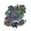
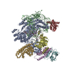
| Entry | Database: PDB / ID: 6fos | |||||||||
|---|---|---|---|---|---|---|---|---|---|---|
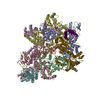
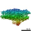
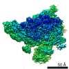
| Title | Cyanidioschyzon merolae photosystem I | |||||||||
 Components Components |
| |||||||||
 Keywords Keywords |  PHOTOSYNTHESIS / membrane protein / photosynthetic complex / reaction center / photosystem PHOTOSYNTHESIS / membrane protein / photosynthetic complex / reaction center / photosystem | |||||||||
| Function / homology |  Function and homology informationthylakoid membrane / photosynthesis, light harvesting / photosystem I reaction center / photosystem I / thylakoid / photosynthetic electron transport in photosystem I / photosystem I / plastid / chlorophyll binding / chloroplast thylakoid membrane ...thylakoid membrane / photosynthesis, light harvesting / photosystem I reaction center / photosystem I / thylakoid / photosynthetic electron transport in photosystem I / photosystem I / plastid / chlorophyll binding / chloroplast thylakoid membrane / photosynthesis / chloroplast / 4 iron, 4 sulfur cluster binding / electron transfer activity / magnesium ion binding / membrane / metal ion binding Function and homology informationthylakoid membrane / photosynthesis, light harvesting / photosystem I reaction center / photosystem I / thylakoid / photosynthetic electron transport in photosystem I / photosystem I / plastid / chlorophyll binding / chloroplast thylakoid membrane ...thylakoid membrane / photosynthesis, light harvesting / photosystem I reaction center / photosystem I / thylakoid / photosynthetic electron transport in photosystem I / photosystem I / plastid / chlorophyll binding / chloroplast thylakoid membrane / photosynthesis / chloroplast / 4 iron, 4 sulfur cluster binding / electron transfer activity / magnesium ion binding / membrane / metal ion bindingSimilarity search - Function | |||||||||
| Biological species |  Cyanidioschyzon merolae (eukaryote) Cyanidioschyzon merolae (eukaryote) | |||||||||
| Method | X-RAY DIFFRACTION / SYNCHROTRON / SAD / Resolution: 4 Å | |||||||||
 Authors Authors | Nelson, N. / Hippler, M. / Antoshvili, M. / Caspy, I. | |||||||||
| Funding support |  Belgium, Belgium,  Israel, 2items Israel, 2items
| |||||||||
 Citation Citation | Journal: Photosyn. Res. / Year: 2019 Title: Structure and function of photosystem I in Cyanidioschyzon merolae. Authors: Antoshvili, M. / Caspy, I. / Hippler, M. / Nelson, N. | |||||||||
| History |
|
- Structure visualization
| Structure viewer | Molecule: MolmilJmol/JSmol |
|---|
- Downloads & links
-Download
| PDBx/mmCIF format | 6fos.cif.gz | 694.6 KB | Display | PDBx/mmCIF format |
|---|---|---|---|---|
| PDB format | pdb6fos.ent.gz | 572.2 KB | Display | PDB format |
| PDBx/mmJSON format | 6fos.json.gz | Tree view | PDBx/mmJSON format | |
| Others |  Other downloads Other downloads |
-Validation report
| Arichive directory | https://data.pdbj.org/pub/pdb/validation_reports/fo/6fosftp://data.pdbj.org/pub/pdb/validation_reports/fo/6fos | HTTPS FTP |
|---|
-Related structure data
| Similar structure data |
|---|

-Links
 PDBj
PDBj


- Assembly
| Deposited unit | 
| ||||||||
|---|---|---|---|---|---|---|---|---|---|
| 1 |
| ||||||||
| Unit cell |
|
-Components
-Protein , 3 types, 4 molecules 234O
| #1: Protein | Mass: 24270.199 Da / Num. of mol.: 2 / Source method: isolated from a natural source Source: (natural) Cyanidioschyzon merolae (strain 10D) (eukaryote)Strain: 10D / References: UniProt: M1UU36 #2: Protein | | Mass: 23987.664 Da / Num. of mol.: 1 / Source method: isolated from a natural source Source: (natural) Cyanidioschyzon merolae (strain 10D) (eukaryote)Strain: 10D / References: UniProt: M1VKK5 #14: Protein | | Mass: 10625.323 Da / Num. of mol.: 1 / Source method: isolated from a natural source Source: (natural) Cyanidioschyzon merolae (strain 10D) (eukaryote)Strain: 10D / References: UniProt: M1VFJ4 |
|---|
-Photosystem I P700 chlorophyll a apoprotein ... , 3 types, 3 molecules ABD
| #3: Protein | / PSI-A / PsaA Mass: 81829.367 Da / Num. of mol.: 1 / Source method: isolated from a natural source Source: (natural) Cyanidioschyzon merolae (strain 10D) (eukaryote)Strain: 10D / References: UniProt: Q85FY7, photosystem I |
|---|---|
| #4: Protein | / PSI-B / PsaB Mass: 81301.820 Da / Num. of mol.: 1 / Source method: isolated from a natural source Source: (natural) Cyanidioschyzon merolae (strain 10D) (eukaryote)Strain: 10D / References: UniProt: Q85FY6, photosystem I |
| #6: Protein | Mass: 13896.780 Da / Num. of mol.: 1 / Source method: isolated from a natural source Source: (natural) Cyanidioschyzon merolae (strain 10D) (eukaryote)Strain: 10D / References: UniProt: Q85FY0 |
-Photosystem I iron-sulfur ... , 2 types, 2 molecules CE
| #5: Protein | / 9 kDa polypeptide / PSI-C / Photosystem I subunit VII / PsaC Mass: 8691.076 Da / Num. of mol.: 1 / Source method: isolated from a natural source Source: (natural) Cyanidioschyzon merolae (strain 10D) (eukaryote)Strain: 10D / References: UniProt: Q85G47, photosystem I |
|---|---|
| #7: Protein | Mass: 7890.087 Da / Num. of mol.: 1 / Source method: isolated from a natural source Source: (natural) Cyanidioschyzon merolae (strain 10D) (eukaryote)Strain: 10D / References: UniProt: Q85FZ1 |
-Photosystem I reaction center subunit ... , 6 types, 6 molecules FIJKLM
| #8: Protein | Mass: 18055.459 Da / Num. of mol.: 1 / Source method: isolated from a natural source Source: (natural) Cyanidioschyzon merolae (strain 10D) (eukaryote)Strain: 10D / References: UniProt: Q85FS9 |
|---|---|
| #9: Protein/peptide | Mass: 3408.184 Da / Num. of mol.: 1 / Source method: isolated from a natural source Source: (natural) Cyanidioschyzon merolae (strain 10D) (eukaryote)Strain: 10D / References: UniProt: Q85FQ6 |
| #10: Protein/peptide | / PSI-J Mass: 4410.245 Da / Num. of mol.: 1 / Source method: isolated from a natural source Source: (natural) Cyanidioschyzon merolae (strain 10D) (eukaryote)Strain: 10D / References: UniProt: Q85FS8 |
| #11: Protein/peptide | Mass: 4820.790 Da / Num. of mol.: 1 / Source method: isolated from a natural source Source: (natural) Cyanidioschyzon merolae (strain 10D) (eukaryote)Strain: 10D / References: UniProt: Q85G51 |
| #12: Protein | / PSI subunit V / PSI-L Mass: 15157.522 Da / Num. of mol.: 1 / Source method: isolated from a natural source Source: (natural) Cyanidioschyzon merolae (strain 10D) (eukaryote)Strain: 10D / References: UniProt: Q85FP8 |
| #13: Protein/peptide | / PSI-M Mass: 3139.878 Da / Num. of mol.: 1 / Source method: isolated from a natural source Source: (natural) Cyanidioschyzon merolae (strain 10D) (eukaryote)Strain: 10D / References: UniProt: Q85G73 |
-Non-polymers , 4 types, 147 molecules 


| #15: Chemical | ChemComp-CLA / Chlorophyll a Mass: 893.489 Da / Num. of mol.: 134 / Source method: obtained synthetically / Formula: C55H72MgN4O5 Mass: 893.489 Da / Num. of mol.: 134 / Source method: obtained synthetically / Formula: C55H72MgN4O5#16: Chemical | Phytomenadione Mass: 450.696 Da / Num. of mol.: 2 / Source method: obtained synthetically / Formula: C31H46O2 Mass: 450.696 Da / Num. of mol.: 2 / Source method: obtained synthetically / Formula: C31H46O2#17: Chemical | Iron–sulfur cluster Mass: 351.640 Da / Num. of mol.: 3 / Source method: obtained synthetically / Formula: Fe4S4 Mass: 351.640 Da / Num. of mol.: 3 / Source method: obtained synthetically / Formula: Fe4S4#18: Chemical | ChemComp-BCR / Β-Carotene Mass: 536.873 Da / Num. of mol.: 8 / Source method: obtained synthetically / Formula: C40H56 Mass: 536.873 Da / Num. of mol.: 8 / Source method: obtained synthetically / Formula: C40H56 |
|---|
-Experimental details
-Experiment
| Experiment | Method: X-RAY DIFFRACTION / Number of used crystals: 1 |
|---|
- Sample preparation
| Crystal | Density Matthews: 5.08 Å3/Da / Density % sol: 75.76 % |
|---|---|
| Crystal grow | Temperature: 277 K / Method: vapor diffusion, sitting drop Details: 80mM MgCl2 , 50mM Citric acid, 4-7.5% PEG 4000, 0.025% alpha-DM, pH 4.8-5.0 or 80mM MgCl2 , 50mM Cacodylate, 4-7.5% PEG 4000, 0.025% alpha-DM, pH 5-6 |
-Data collection
| Diffraction | Mean temperature: 100 K |
|---|---|
| Diffraction source | Source: SYNCHROTRON / Site: SLS  / Beamline: X06SA / Wavelength: 0.999 Å / Beamline: X06SA / Wavelength: 0.999 Å |
| Detector | Type: DECTRIS EIGER X 16M / Detector: PIXEL / Date: Jul 8, 2017 |
| Radiation | Protocol: SINGLE WAVELENGTH / Monochromatic (M) / Laue (L): M / Scattering type: x-ray |
| Radiation wavelength | Wavelength: 0.999 Å / Relative weight: 1 |
| Reflection | Resolution: 4→49 Å / Num. obs: 41951 / % possible obs: 80.89 % / Redundancy: 12 % / Rpim(I) all: 0.036 / Net I/σ(I): 4 |
| Reflection shell | Resolution: 4→4.99 Å / Rpim(I) all: 1.236 |
- Processing
| Software |
| |||||||||||||||||||||||||||||||||||||||||||||||||
|---|---|---|---|---|---|---|---|---|---|---|---|---|---|---|---|---|---|---|---|---|---|---|---|---|---|---|---|---|---|---|---|---|---|---|---|---|---|---|---|---|---|---|---|---|---|---|---|---|---|---|
| Refinement | Method to determine structure: SAD / Resolution: 4→48.53 Å / SU ML: 0.93 / Cross valid method: FREE R-VALUE / σ(F): 1.36 / Phase error: 55.96 / Stereochemistry target values: ML
| |||||||||||||||||||||||||||||||||||||||||||||||||
| Solvent computation | Shrinkage radii: 0.9 Å / VDW probe radii: 1.11 Å / Solvent model: FLAT BULK SOLVENT MODEL | |||||||||||||||||||||||||||||||||||||||||||||||||
| Refinement step | Cycle: LAST / Resolution: 4→48.53 Å
| |||||||||||||||||||||||||||||||||||||||||||||||||
| Refine LS restraints |
| |||||||||||||||||||||||||||||||||||||||||||||||||
| LS refinement shell |
|
