 Movie
Movie Controller

[English] 日本語
 Yorodumi
- PDB-6fne: Structure of human Brag2 (Sec7-PH domains) with the inhibitor Bra... -
+ Open data

- Basic information
| Entry | Database: PDB / ID: 6fne | ||||||
|---|---|---|---|---|---|---|---|
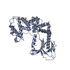
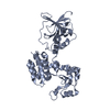
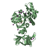
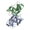
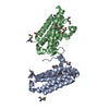
| Title | Structure of human Brag2 (Sec7-PH domains) with the inhibitor Bragsin bound to the PH domain | ||||||
 Components Components | IQ motif and SEC7 domain-containing protein 1 | ||||||
 Keywords Keywords |  HYDROLASE / ARF GEF HYDROLASE / ARF GEF | ||||||
| Function / homology |  Function and homology information Function and homology informationpositive regulation of keratinocyte migration / dendritic spine development / positive regulation of focal adhesion disassembly / regulation of ARF protein signal transduction / guanyl-nucleotide exchange factor activity / synaptic vesicle / actin cytoskeleton organization / postsynaptic density / intracellular membrane-bounded organelle / lipid binding ...positive regulation of keratinocyte migration / dendritic spine development / positive regulation of focal adhesion disassembly / regulation of ARF protein signal transduction / guanyl-nucleotide exchange factor activity / synaptic vesicle / actin cytoskeleton organization / postsynaptic density / intracellular membrane-bounded organelle / lipid binding / nucleolus / membraneSimilarity search - Function | ||||||
| Biological species |  Homo sapiens (human) Homo sapiens (human) | ||||||
| Method | X-RAY DIFFRACTION / SYNCHROTRON / MOLECULAR REPLACEMENT / Resolution: 2.501 Å | ||||||
 Authors Authors | Nawrotek, A. / Zeghouf, M. / Cherfils, J. | ||||||
| Funding support |  France, 1items France, 1items
| ||||||
 Citation Citation | Journal: Nat.Chem.Biol. / Year: 2019 Title: PH-domain-binding inhibitors of nucleotide exchange factor BRAG2 disrupt Arf GTPase signaling. Authors: Nawrotek, A. / Benabdi, S. / Niyomchon, S. / Kryszke, M.H. / Ginestier, C. / Caneque, T. / Tepshi, L. / Mariani, A. / St Onge, R.P. / Giaever, G. / Nislow, C. / Charafe-Jauffret, E. / ...Authors: Nawrotek, A. / Benabdi, S. / Niyomchon, S. / Kryszke, M.H. / Ginestier, C. / Caneque, T. / Tepshi, L. / Mariani, A. / St Onge, R.P. / Giaever, G. / Nislow, C. / Charafe-Jauffret, E. / Rodriguez, R. / Zeghouf, M. / Cherfils, J. | ||||||
| History |
|
- Structure visualization
| Structure viewer | Molecule: MolmilJmol/JSmol |
|---|
- Downloads & links
-Download
| PDBx/mmCIF format | 6fne.cif.gz | 155.3 KB | Display | PDBx/mmCIF format |
|---|---|---|---|---|
| PDB format | pdb6fne.ent.gz | 119.9 KB | Display | PDB format |
| PDBx/mmJSON format | 6fne.json.gz | Tree view | PDBx/mmJSON format | |
| Others |  Other downloads Other downloads |
-Validation report
| Arichive directory | https://data.pdbj.org/pub/pdb/validation_reports/fn/6fneftp://data.pdbj.org/pub/pdb/validation_reports/fn/6fne | HTTPS FTP |
|---|
-Related structure data
| Related structure data |  5nlyS S: Starting model for refinement |
|---|---|
| Similar structure data |


-Links
 PDBj
PDBj

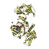
- Assembly
| Deposited unit | 
| ||||||||
|---|---|---|---|---|---|---|---|---|---|
| 1 | 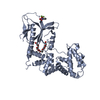
| ||||||||
| 2 | 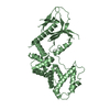
| ||||||||
| Unit cell |
|
-Components
| #1: Protein | Mass: 47392.555 Da / Num. of mol.: 2 Source method: isolated from a genetically manipulated source Source: (gene. exp.) Homo sapiens (human) / Gene: IQSEC1, ARFGEP100, BRAG2, KIAA0763 / Production host:  Escherichia coli (E. coli) / References: UniProt: Q6DN90 Escherichia coli (E. coli) / References: UniProt: Q6DN90#2: Chemical | ChemComp-DY5 / ( |   Mass: 275.181 Da / Num. of mol.: 1 / Source method: obtained synthetically / Formula: C11H8F3NO4 / Feature type: SUBJECT OF INVESTIGATION Mass: 275.181 Da / Num. of mol.: 1 / Source method: obtained synthetically / Formula: C11H8F3NO4 / Feature type: SUBJECT OF INVESTIGATION#3: Chemical | ChemComp-2PE / | Polyethylene glycol  Mass: 414.488 Da / Num. of mol.: 1 / Source method: isolated from a natural source / Formula: C18H38O10 / Comment: precipitant*YM Mass: 414.488 Da / Num. of mol.: 1 / Source method: isolated from a natural source / Formula: C18H38O10 / Comment: precipitant*YM#4: Water | ChemComp-HOH / | Water Mass: 18.015 Da / Num. of mol.: 18 / Source method: isolated from a natural source / Formula: H2O Mass: 18.015 Da / Num. of mol.: 18 / Source method: isolated from a natural source / Formula: H2O |
|---|
-Experimental details
-Experiment
| Experiment | Method: X-RAY DIFFRACTION / Number of used crystals: 1 |
|---|
- Sample preparation
| Crystal | Density Matthews: 2.52 Å3/Da / Density % sol: 51.13 % |
|---|---|
| Crystal grow | Temperature: 293 K / Method: vapor diffusion / Details: 18% PEG 20000, 0.1 M Tris pH 8.5 |
-Data collection
| Diffraction | Mean temperature: 80 K |
|---|---|
| Diffraction source | Source: SYNCHROTRON / Site: SOLEIL / Beamline: PROXIMA 2 / Wavelength: 0.9801 Å |
| Detector | Type: DECTRIS EIGER X 16M / Detector: PIXEL / Date: Mar 20, 2016 |
| Radiation | Protocol: SINGLE WAVELENGTH / Monochromatic (M) / Laue (L): M / Scattering type: x-ray |
| Radiation wavelength | Wavelength: 0.9801 Å / Relative weight: 1 |
| Reflection | Resolution: 2.5→65.9 Å / Num. all: 286171 / Num. obs: 21954 / % possible obs: 93.9 % / Redundancy: 13 % / Biso Wilson estimate: 70.64 Å2 / CC1/2: 0.997 / Rpim(I) all: 0.05 / Rsym value: 0.18 / Net I/σ(I): 9.9 |
| Reflection shell | Resolution: 2.5→2.78 Å / Num. measured obs: 11487 / Num. unique all: 1099 / Num. unique obs: 11487 / Rpim(I) all: 0.46 / % possible all: 70 |
- Processing
| Software |
| |||||||||||||||||||||||||||||||||||||||||||||||||||||||||||||||
|---|---|---|---|---|---|---|---|---|---|---|---|---|---|---|---|---|---|---|---|---|---|---|---|---|---|---|---|---|---|---|---|---|---|---|---|---|---|---|---|---|---|---|---|---|---|---|---|---|---|---|---|---|---|---|---|---|---|---|---|---|---|---|---|---|
| Refinement | Method to determine structure: MOLECULAR REPLACEMENT Starting model: 5NLY Resolution: 2.501→63.33 Å / SU ML: 0.35 / Cross valid method: THROUGHOUT / σ(F): 1.36 / Phase error: 35.53
| |||||||||||||||||||||||||||||||||||||||||||||||||||||||||||||||
| Solvent computation | Shrinkage radii: 0.9 Å / VDW probe radii: 1.11 Å | |||||||||||||||||||||||||||||||||||||||||||||||||||||||||||||||
| Refinement step | Cycle: LAST / Resolution: 2.501→63.33 Å
| |||||||||||||||||||||||||||||||||||||||||||||||||||||||||||||||
| Refine LS restraints |
| |||||||||||||||||||||||||||||||||||||||||||||||||||||||||||||||
| LS refinement shell |
|
