 Movie
Movie Controller

[English] 日本語
 Yorodumi
- PDB-6fnb: Mono- and bivalent 14-3-3 inhibitors for characterizing supramole... -
+ Open data

- Basic information
| Entry | Database: PDB / ID: 6fnb | ||||||
|---|---|---|---|---|---|---|---|
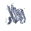
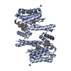
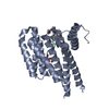
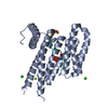
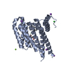
| Title | Mono- and bivalent 14-3-3 inhibitors for characterizing supramolecular lysine-PEG interactions in proteins | ||||||
 Components Components | 14-3-3 protein zeta/delta | ||||||
 Keywords Keywords |  PROTEIN BINDING / Inhibition / Mono- and bivalent 14-3-3 inhibitors / Supramolecular lysine-PEG PROTEIN BINDING / Inhibition / Mono- and bivalent 14-3-3 inhibitors / Supramolecular lysine-PEG | ||||||
| Function / homology |  Function and homology information Function and homology informationGolgi reassembly / regulation of synapse maturation / NOTCH4 Activation and Transmission of Signal to the Nucleus / establishment of Golgi localization / Rap1 signalling / negative regulation of protein localization to nucleus / KSRP (KHSRP) binds and destabilizes mRNA / GP1b-IX-V activation signalling / Regulation of localization of FOXO transcription factors / Interleukin-3, Interleukin-5 and GM-CSF signaling ...Golgi reassembly / regulation of synapse maturation / NOTCH4 Activation and Transmission of Signal to the Nucleus / establishment of Golgi localization / Rap1 signalling / negative regulation of protein localization to nucleus / KSRP (KHSRP) binds and destabilizes mRNA / GP1b-IX-V activation signalling / Regulation of localization of FOXO transcription factors / Interleukin-3, Interleukin-5 and GM-CSF signaling / phosphoserine residue binding / Activation of BAD and translocation to mitochondria / SARS-CoV-2 targets host intracellular signalling and regulatory pathways / cellular response to glucose starvation / Chk1/Chk2(Cds1) mediated inactivation of Cyclin B:Cdk1 complex / SARS-CoV-1 targets host intracellular signalling and regulatory pathways / RHO GTPases activate PKNs / negative regulation of TORC1 signaling / negative regulation of innate immune response / protein sequestering activity / regulation of ERK1 and ERK2 cascade / Translocation of SLC2A4 (GLUT4) to the plasma membrane / Deactivation of the beta-catenin transactivating complex / TP53 Regulates Metabolic Genes / Negative regulation of NOTCH4 signaling / melanosome / DNA-binding transcription factor binding / vesicle / transmembrane transporter binding / blood microparticle / cadherin binding / protein phosphorylation / focal adhesion / glutamatergic synapse / ubiquitin protein ligase binding / negative regulation of apoptotic process / protein kinase binding / negative regulation of transcription by RNA polymerase II / signal transduction / extracellular space / RNA binding / extracellular exosome / nucleoplasm / identical protein binding / nucleus / cytosol / cytoplasmSimilarity search - Function | ||||||
| Biological species |  Homo sapiens (human) Homo sapiens (human) | ||||||
| Method | X-RAY DIFFRACTION / SYNCHROTRON / MOLECULAR REPLACEMENT / molecular replacement / Resolution: 2.3 Å | ||||||
 Authors Authors | Bier, D. / Ottmann, C. | ||||||
| Funding support |  Germany, 1items Germany, 1items
| ||||||
 Citation Citation | Journal: Chemistry / Year: 2018 Title: Mono- and Bivalent 14-3-3 Inhibitors for Characterizing Supramolecular "Lysine Wrapping" of Oligoethylene Glycol (OEG) Moieties in Proteins. Authors: Yilmaz, E. / Bier, D. / Guillory, X. / Briels, J. / Ruiz-Blanco, Y.B. / Sanchez-Garcia, E. / Ottmann, C. / Kaiser, M. | ||||||
| History |
|
- Structure visualization
| Structure viewer | Molecule: MolmilJmol/JSmol |
|---|
- Downloads & links
-Download
| PDBx/mmCIF format | 6fnb.cif.gz | 112.4 KB | Display | PDBx/mmCIF format |
|---|---|---|---|---|
| PDB format | pdb6fnb.ent.gz | 82.2 KB | Display | PDB format |
| PDBx/mmJSON format | 6fnb.json.gz | Tree view | PDBx/mmJSON format | |
| Others |  Other downloads Other downloads |
-Validation report
| Arichive directory | https://data.pdbj.org/pub/pdb/validation_reports/fn/6fnbftp://data.pdbj.org/pub/pdb/validation_reports/fn/6fnb | HTTPS FTP |
|---|
-Related structure data
| Related structure data |  6fn9C  6fnaC  6fncC  3nkxS S: Starting model for refinement C: citing same article ( |
|---|---|
| Similar structure data |


-Links
 PDBj
PDBj


- Assembly
| Deposited unit | 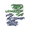
| ||||||||
|---|---|---|---|---|---|---|---|---|---|
| 1 |
| ||||||||
| Unit cell |
|
-Components
-Protein , 1 types, 2 molecules AB
| #1: Protein | Mass: 26316.764 Da / Num. of mol.: 2 Source method: isolated from a genetically manipulated source Source: (gene. exp.) Homo sapiens (human) / Gene: YWHAZ / Production host:  Escherichia coli (E. coli) / References: UniProt: P63104 Escherichia coli (E. coli) / References: UniProt: P63104 |
|---|
-Non-polymers , 6 types, 191 molecules 


| #2: Chemical | Benzoic acid Mass: 122.121 Da / Num. of mol.: 2 / Source method: obtained synthetically / Formula: C7H6O2 Mass: 122.121 Da / Num. of mol.: 2 / Source method: obtained synthetically / Formula: C7H6O2#3: Chemical | Glycerol Mass: 92.094 Da / Num. of mol.: 3 / Source method: obtained synthetically / Formula: C3H8O3 Mass: 92.094 Da / Num. of mol.: 3 / Source method: obtained synthetically / Formula: C3H8O3#4: Chemical |  Mass: 40.078 Da / Num. of mol.: 2 / Source method: obtained synthetically / Formula: Ca Mass: 40.078 Da / Num. of mol.: 2 / Source method: obtained synthetically / Formula: Ca#5: Chemical | ChemComp-CL / Chloride Mass: 35.453 Da / Num. of mol.: 4 / Source method: obtained synthetically / Formula: Cl Mass: 35.453 Da / Num. of mol.: 4 / Source method: obtained synthetically / Formula: Cl#6: Chemical |  Mass: 1291.273 Da / Num. of mol.: 2 / Source method: obtained synthetically / Formula: C59H84N6O22P2 Mass: 1291.273 Da / Num. of mol.: 2 / Source method: obtained synthetically / Formula: C59H84N6O22P2#7: Water | ChemComp-HOH / | WaterMass: 18.015 Da / Num. of mol.: 178 / Source method: isolated from a natural source / Formula: H2O |
|---|
-Experimental details
-Experiment
| Experiment | Method: X-RAY DIFFRACTION / Number of used crystals: 1 |
|---|
- Sample preparation
| Crystal | Density Matthews: 3.98 Å3/Da / Density % sol: 69.12 % |
|---|---|
| Crystal grow | Temperature: 277.15 K / Method: vapor diffusion, sitting drop Details: 0.09 M HEPES sodium salt pH 7.5 1.26 M tri-Sodium citrate 10 %(v/v) Glycerol |
-Data collection
| Diffraction | Mean temperature: 100 K | ||||||||||||||||||||||||||||||||||||||||||||||||||||||||||||||||||||||||||||||||||||||||||||||||||||||||||||||||||||||||||||||||||
|---|---|---|---|---|---|---|---|---|---|---|---|---|---|---|---|---|---|---|---|---|---|---|---|---|---|---|---|---|---|---|---|---|---|---|---|---|---|---|---|---|---|---|---|---|---|---|---|---|---|---|---|---|---|---|---|---|---|---|---|---|---|---|---|---|---|---|---|---|---|---|---|---|---|---|---|---|---|---|---|---|---|---|---|---|---|---|---|---|---|---|---|---|---|---|---|---|---|---|---|---|---|---|---|---|---|---|---|---|---|---|---|---|---|---|---|---|---|---|---|---|---|---|---|---|---|---|---|---|---|---|---|
| Diffraction source | Source: SYNCHROTRON / Site: SLS  / Beamline: X10SA / Wavelength: 1 Å / Beamline: X10SA / Wavelength: 1 Å | ||||||||||||||||||||||||||||||||||||||||||||||||||||||||||||||||||||||||||||||||||||||||||||||||||||||||||||||||||||||||||||||||||
| Detector | Type: DECTRIS PILATUS 6M / Detector: PIXEL / Date: Sep 20, 2016 | ||||||||||||||||||||||||||||||||||||||||||||||||||||||||||||||||||||||||||||||||||||||||||||||||||||||||||||||||||||||||||||||||||
| Radiation | Protocol: SINGLE WAVELENGTH / Monochromatic (M) / Laue (L): M / Scattering type: x-ray | ||||||||||||||||||||||||||||||||||||||||||||||||||||||||||||||||||||||||||||||||||||||||||||||||||||||||||||||||||||||||||||||||||
| Radiation wavelength | Wavelength: 1 Å / Relative weight: 1 | ||||||||||||||||||||||||||||||||||||||||||||||||||||||||||||||||||||||||||||||||||||||||||||||||||||||||||||||||||||||||||||||||||
| Reflection | Resolution: 2.3→46.98 Å / Num. obs: 38130 / % possible obs: 100 % / Redundancy: 13.252 % / Biso Wilson estimate: 60.572 Å2 / CC1/2: 0.996 / Rmerge(I) obs: 0.128 / Rrim(I) all: 0.134 / Χ2: 0.95 / Net I/σ(I): 12.09 / Num. measured all: 505296 / Scaling rejects: 27 | ||||||||||||||||||||||||||||||||||||||||||||||||||||||||||||||||||||||||||||||||||||||||||||||||||||||||||||||||||||||||||||||||||
| Reflection shell | Diffraction-ID: 1
|
-Phasing
| Phasing | Method: molecular replacement | |||||||||
|---|---|---|---|---|---|---|---|---|---|---|
| Phasing MR | Model details: Phaser MODE: MR_AUTO
|
- Processing
| Software |
| ||||||||||||||||||||||||||||||||||||||||||||||||||||||||||||
|---|---|---|---|---|---|---|---|---|---|---|---|---|---|---|---|---|---|---|---|---|---|---|---|---|---|---|---|---|---|---|---|---|---|---|---|---|---|---|---|---|---|---|---|---|---|---|---|---|---|---|---|---|---|---|---|---|---|---|---|---|---|
| Refinement | Method to determine structure: MOLECULAR REPLACEMENT Starting model: 3NKX Resolution: 2.3→46.98 Å / Cor.coef. Fo:Fc: 0.956 / Cor.coef. Fo:Fc free: 0.946 / SU B: 7.254 / SU ML: 0.163 / SU R Cruickshank DPI: 0.2152 / Cross valid method: THROUGHOUT / σ(F): 0 / ESU R: 0.215 / ESU R Free: 0.189 Details: HYDROGENS HAVE BEEN ADDED IN THE RIDING POSITIONS U VALUES : REFINED INDIVIDUALLY
| ||||||||||||||||||||||||||||||||||||||||||||||||||||||||||||
| Solvent computation | Ion probe radii: 0.8 Å / Shrinkage radii: 0.8 Å / VDW probe radii: 1.2 Å | ||||||||||||||||||||||||||||||||||||||||||||||||||||||||||||
| Displacement parameters | Biso max: 137.39 Å2 / Biso mean: 60.153 Å2 / Biso min: 36.23 Å2
| ||||||||||||||||||||||||||||||||||||||||||||||||||||||||||||
| Refinement step | Cycle: final / Resolution: 2.3→46.98 Å
| ||||||||||||||||||||||||||||||||||||||||||||||||||||||||||||
| Refine LS restraints |
| ||||||||||||||||||||||||||||||||||||||||||||||||||||||||||||
| LS refinement shell | Resolution: 2.3→2.36 Å / Rfactor Rfree error: 0 / Total num. of bins used: 20
|
