Protocol: SINGLE WAVELENGTH / Monochromatic (M) / Laue (L): M / Scattering type: x-ray
Radiation wavelength
Wavelength: 0.992 Å / Relative weight: 1
Reflection
Resolution: 2.3→29.8 Å / Num. obs: 22003 / % possible obs: 96.6 % / Redundancy: 1.53 % / Biso Wilson estimate: 19.75 Å2 / Rmerge(I) obs: 0.07 / Net I/σ(I): 7.15
Reflection shell
Resolution: 2.3→2.4 Å
-
Processing
Software
Name
Version
Classification
REFMAC
5.8.0123
refinement
XDS
datareduction
SCALA
datascaling
MOLREP
phasing
Refinement
Resolution: 2.3→29.8 Å / Cor.coef. Fo:Fc: 0.946 / Cor.coef. Fo:Fc free: 0.9 / SU B: 7.14 / SU ML: 0.175 / Cross valid method: THROUGHOUT / ESU R: 0.407 / ESU R Free: 0.241 / Details: HYDROGENS HAVE BEEN ADDED IN THE RIDING POSITIONS
Rfactor
Num. reflection
% reflection
Selection details
Rfree
0.22753
682
3.1 %
RANDOM
Rwork
0.16778
-
-
-
obs
0.16963
21265
96.53 %
-
Solvent computation
Ion probe radii: 0.8 Å / Shrinkage radii: 0.8 Å / VDW probe radii: 1.2 Å
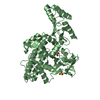
 Movie
Movie Controller

 Yorodumi
 Open data
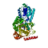
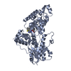
 Basic information
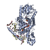
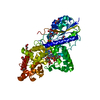
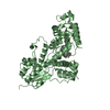
 Components
 Keywords
 FLAVOPROTEIN /
  Function and homology information

 Authors
 Germany, 1items
  Citation
 Structure visualization
 Downloads & links
 Other downloads
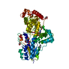


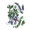


 PDBj
PDBj
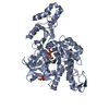
 Assembly
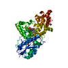


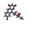


 Mass: 785.550 Da / Num. of mol.: 1 / Source method: obtained synthetically / Formula: C27H33N9O15P2 / Comment: FAD*YM
 Mass: 363.322 Da / Num. of mol.: 1 / Source method: obtained synthetically / Formula: C16H17N3O7
 Mass: 92.094 Da / Num. of mol.: 3 / Source method: obtained synthetically / Formula: C3H8O3
 Mass: 94.971 Da / Num. of mol.: 1 / Source method: obtained synthetically / Formula: PO4
 Mass: 35.453 Da / Num. of mol.: 1 / Source method: obtained synthetically / Formula: Cl
 Sample preparation
 / Beamline: ID23-2 / Wavelength: 0.992 Å
 Processing