 Movie
Movie Controller

[English] 日本語
 Yorodumi
- PDB-6fm3: Deoxyguanylosuccinate synthase (DgsS) structure with ADP at 1.9 A... -
+ Open data

- Basic information
| Entry | Database: PDB / ID: 6fm3 | ||||||
|---|---|---|---|---|---|---|---|
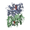
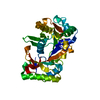
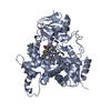
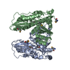
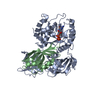
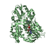
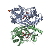
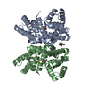
| Title | Deoxyguanylosuccinate synthase (DgsS) structure with ADP at 1.9 Angstrom resolution | ||||||
 Components Components | Adenylosuccinate synthetase Adenylosuccinate synthase Adenylosuccinate synthase | ||||||
 Keywords Keywords | BIOSYNTHETIC PROTEIN / 2 / 6-diaminopurine / phage phiVC8 / Synthetase | ||||||
| Function / homology |  Function and homology information Function and homology information2-amino-2'-deoxyadenylo-succinate synthase / adenylosuccinate synthase activity / purine nucleotide biosynthetic process / magnesium ion binding / ATP bindingSimilarity search - Function | ||||||
| Biological species |  Vibrio phage phiVC8 (virus) Vibrio phage phiVC8 (virus) | ||||||
| Method | X-RAY DIFFRACTION / Resolution: 1.95 Å | ||||||
 Authors Authors | Sleiman, D. / Loc'h, J. / Haouz, A. / Kaminski, P.A. | ||||||
 Citation Citation | Journal: To Be Published Title: Deoxyguanylosuccinate synthase (DgsS) quaternary structure with ATP0, dGMP, Magnesium at 2.3 Angstrom resolution Authors: Sleiman, D. / Loc'h, J. / Haouz, A. / Kaminski, P.A. | ||||||
| History |
|
- Structure visualization
| Structure viewer | Molecule: MolmilJmol/JSmol |
|---|
- Downloads & links
-Download
| PDBx/mmCIF format | 6fm3.cif.gz | 142.6 KB | Display | PDBx/mmCIF format |
|---|---|---|---|---|
| PDB format | pdb6fm3.ent.gz | 108.5 KB | Display | PDB format |
| PDBx/mmJSON format | 6fm3.json.gz | Tree view | PDBx/mmJSON format | |
| Others |  Other downloads Other downloads |
-Validation report
| Arichive directory | https://data.pdbj.org/pub/pdb/validation_reports/fm/6fm3ftp://data.pdbj.org/pub/pdb/validation_reports/fm/6fm3 | HTTPS FTP |
|---|
-Related structure data
| Related structure data | |
|---|---|
| Similar structure data |


-Links
 PDBj
PDBj- Assembly
| Deposited unit | 
| ||||||||
|---|---|---|---|---|---|---|---|---|---|
| 1 | 
| ||||||||
| Unit cell |
| ||||||||
| Components on special symmetry positions |
|
-Components
| #1: Protein | Adenylosuccinate synthase Mass: 40392.996 Da / Num. of mol.: 1 Source method: isolated from a genetically manipulated source Source: (gene. exp.) Vibrio phage phiVC8 (virus) / Gene: phiVC8_p27 / Production host:  Escherichia coli BL21(DE3) (bacteria) / References: UniProt: G3FFN6 Escherichia coli BL21(DE3) (bacteria) / References: UniProt: G3FFN6 |
|---|---|
| #2: Chemical | ChemComp-ADP / Adenosine diphosphate  Mass: 427.201 Da / Num. of mol.: 1 / Source method: obtained synthetically / Formula: C10H15N5O10P2 / Feature type: SUBJECT OF INVESTIGATION / Comment: ADP, energy-carrying molecule*YM Mass: 427.201 Da / Num. of mol.: 1 / Source method: obtained synthetically / Formula: C10H15N5O10P2 / Feature type: SUBJECT OF INVESTIGATION / Comment: ADP, energy-carrying molecule*YM |
| #3: Chemical | ChemComp-CL / Chloride  Mass: 35.453 Da / Num. of mol.: 1 / Source method: obtained synthetically / Formula: Cl Mass: 35.453 Da / Num. of mol.: 1 / Source method: obtained synthetically / Formula: Cl |
| #4: Water | ChemComp-HOH / Water Mass: 18.015 Da / Num. of mol.: 357 / Source method: isolated from a natural source / Formula: H2O Mass: 18.015 Da / Num. of mol.: 357 / Source method: isolated from a natural source / Formula: H2O |
-Experimental details
-Experiment
| Experiment | Method: X-RAY DIFFRACTION / Number of used crystals: 1 |
|---|
- Sample preparation
| Crystal | Density Matthews: 2.25 Å3/Da / Density % sol: 45.27 % |
|---|---|
| Crystal grow | Temperature: 290 K / Method: vapor diffusion, sitting drop / Details: 2M NaCl 12%w/v PEG 6K |
-Data collection
| Diffraction | Mean temperature: 100 K |
|---|---|
| Diffraction source | Source: ROTATING ANODE / Type: RIGAKU MICROMAX-007 / Wavelength: 1.54179 Å |
| Detector | Type: MAR scanner 345 mm plate / Detector: IMAGE PLATE / Date: Jan 4, 2017 |
| Radiation | Protocol: SINGLE WAVELENGTH / Monochromatic (M) / Laue (L): M / Scattering type: x-ray |
| Radiation wavelength | Wavelength: 1.54179 Å / Relative weight: 1 |
| Reflection | Resolution: 1.9→48.32 Å / Num. obs: 24769 / % possible obs: 93.7 % / Redundancy: 5.59 % / Biso Wilson estimate: 41.98 Å2 / Net I/σ(I): 8.6 |
| Reflection shell | Resolution: 2.3→2.44 Å |
- Processing
| Software |
| ||||||||||||||||||||||||||||||||||||||||||||||||||||||||||||||||||||||||||||||||||||||||||||||||||||||||||||||||||
|---|---|---|---|---|---|---|---|---|---|---|---|---|---|---|---|---|---|---|---|---|---|---|---|---|---|---|---|---|---|---|---|---|---|---|---|---|---|---|---|---|---|---|---|---|---|---|---|---|---|---|---|---|---|---|---|---|---|---|---|---|---|---|---|---|---|---|---|---|---|---|---|---|---|---|---|---|---|---|---|---|---|---|---|---|---|---|---|---|---|---|---|---|---|---|---|---|---|---|---|---|---|---|---|---|---|---|---|---|---|---|---|---|---|---|---|
| Refinement | Resolution: 1.95→48.32 Å / Cor.coef. Fo:Fc: 0.921 / Cor.coef. Fo:Fc free: 0.903 / SU R Cruickshank DPI: 0.194 / Cross valid method: THROUGHOUT / σ(F): 0 / SU R Blow DPI: 0.216 / SU Rfree Blow DPI: 0.174 / SU Rfree Cruickshank DPI: 0.166
| ||||||||||||||||||||||||||||||||||||||||||||||||||||||||||||||||||||||||||||||||||||||||||||||||||||||||||||||||||
| Displacement parameters | Biso mean: 36.62 Å2
| ||||||||||||||||||||||||||||||||||||||||||||||||||||||||||||||||||||||||||||||||||||||||||||||||||||||||||||||||||
| Refine analyze | Luzzati coordinate error obs: 0.33 Å | ||||||||||||||||||||||||||||||||||||||||||||||||||||||||||||||||||||||||||||||||||||||||||||||||||||||||||||||||||
| Refinement step | Cycle: 1 / Resolution: 1.95→48.32 Å
| ||||||||||||||||||||||||||||||||||||||||||||||||||||||||||||||||||||||||||||||||||||||||||||||||||||||||||||||||||
| Refine LS restraints |
| ||||||||||||||||||||||||||||||||||||||||||||||||||||||||||||||||||||||||||||||||||||||||||||||||||||||||||||||||||
| LS refinement shell | Resolution: 1.95→2.04 Å / Total num. of bins used: 12
| ||||||||||||||||||||||||||||||||||||||||||||||||||||||||||||||||||||||||||||||||||||||||||||||||||||||||||||||||||
| Refinement TLS params. | Method: refined / Origin x: -11.3624 Å / Origin y: -11.4532 Å / Origin z: 21.3026 Å
| ||||||||||||||||||||||||||||||||||||||||||||||||||||||||||||||||||||||||||||||||||||||||||||||||||||||||||||||||||
| Refinement TLS group | Selection details: { A|* } |
