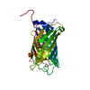
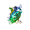
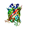
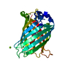
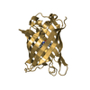
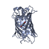
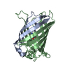
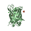
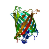
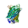
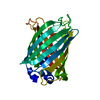
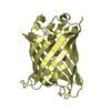
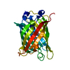
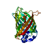
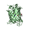
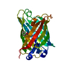
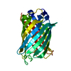
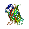
Green Fluorescent Protein / Green fluorescent protein / Green fluorescent protein, GFP / Green fluorescent protein-related / Green fluorescent protein / Green fluorescent protein / Beta Barrel / Mainly Beta Similarity search - Domain/homology
Resolution: 1.79→45.5 Å / Cor.coef. Fo:Fc: 0.943 / Cor.coef. Fo:Fc free: 0.928 / SU B: 3.555 / SU ML: 0.106 / Cross valid method: THROUGHOUT / ESU R: 0.147 / ESU R Free: 0.132 / Stereochemistry target values: MAXIMUM LIKELIHOOD / Details: HYDROGENS HAVE BEEN ADDED IN THE RIDING POSITIONS
Rfactor
Num. reflection
% reflection
Selection details
Rfree
0.23216
1085
5.2 %
RANDOM
Rwork
0.20207
-
-
-
obs
0.20371
19807
99.98 %
-
Solvent computation
Ion probe radii: 0.8 Å / Shrinkage radii: 0.8 Å / VDW probe radii: 1.2 Å / Solvent model: MASK
 Movie
Movie Controller

 Yorodumi
 Open data
 Basic information
 Components
 Keywords
 Function and homology information

 Authors
 Italy, 1items
  Citation
 Structure visualization
 Downloads & links
 Other downloads


 PDBj
PDBj

 Assembly
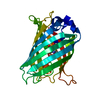

 Mass: 18.015 Da / Num. of mol.: 123 / Source method: isolated from a natural source / Formula: H2O
 Sample preparation
 Processing