 Movie
Movie Controller

[English] 日本語
 Yorodumi
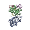
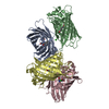
- PDB-6f97: Crystal structure of the V465T mutant of 5-(Hydroxymethyl)furfura... -
+ Open data

- Basic information
| Entry | Database: PDB / ID: 6f97 | ||||||
|---|---|---|---|---|---|---|---|
| Title | Crystal structure of the V465T mutant of 5-(Hydroxymethyl)furfural Oxidase (HMFO) | ||||||
 Components Components | 5-(hydroxymethyl)furfural oxidase | ||||||
 Keywords Keywords |  FLAVOPROTEIN / sec-thiol oxidation / alcohol oxidase / kinetic resolution / biocatalysis / enzyme engineering FLAVOPROTEIN / sec-thiol oxidation / alcohol oxidase / kinetic resolution / biocatalysis / enzyme engineering | ||||||
| Function / homology |  Function and homology information Function and homology information5-(hydroxymethyl)furfural oxidase / Oxidoreductases; Acting on a sulfur group of donors; With oxygen as acceptor / oxidoreductase activity, acting on a sulfur group of donors, oxygen as acceptor / oxidoreductase activity, acting on the CH-OH group of donors, oxygen as acceptor / FAD bindingSimilarity search - Function | ||||||
| Biological species | Methylovorus sp. | ||||||
| Method | X-RAY DIFFRACTION / SYNCHROTRON / MOLECULAR REPLACEMENT / Resolution: 1.9 Å | ||||||
 Authors Authors | Pickl, M. / Swoboda, A. / Romero, E. / Winkler, C.K. / Binda, C. / Mattevi, A. / Faber, K. / Fraaije, M.W. | ||||||
 Citation Citation | Journal: Angew. Chem. Int. Ed. Engl. / Year: 2018 Title: Kinetic Resolution of sec-Thiols by Enantioselective Oxidation with Rationally Engineered 5-(Hydroxymethyl)furfural Oxidase. Authors: Pickl, M. / Swoboda, A. / Romero, E. / Winkler, C.K. / Binda, C. / Mattevi, A. / Faber, K. / Fraaije, M.W. | ||||||
| History |
|
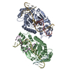
- Structure visualization
| Structure viewer | Molecule: MolmilJmol/JSmol |
|---|
- Downloads & links
-Download
| PDBx/mmCIF format | 6f97.cif.gz | 210.2 KB | Display | PDBx/mmCIF format |
|---|---|---|---|---|
| PDB format | pdb6f97.ent.gz | 167.1 KB | Display | PDB format |
| PDBx/mmJSON format | 6f97.json.gz | Tree view | PDBx/mmJSON format | |
| Others |  Other downloads Other downloads |
-Validation report
| Arichive directory | https://data.pdbj.org/pub/pdb/validation_reports/f9/6f97ftp://data.pdbj.org/pub/pdb/validation_reports/f9/6f97 | HTTPS FTP |
|---|
-Related structure data
| Related structure data |  4udpS S: Starting model for refinement |
|---|---|
| Similar structure data |


-Links
 PDBj
PDBj
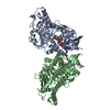
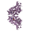
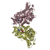
- Assembly
| Deposited unit | 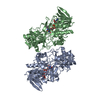
| ||||||||
|---|---|---|---|---|---|---|---|---|---|
| 1 |
| ||||||||
| Unit cell |
|
-Components
| #1: Protein | Mass: 57078.086 Da / Num. of mol.: 2 Source method: isolated from a genetically manipulated source Details: The sequence contains the V465T mutation (with respect to the wild type sequence) which was intentionally introduced by site-directed mutagenesis. Source: (gene. exp.)  Methylovorus sp. (strain MP688) (bacteria) Methylovorus sp. (strain MP688) (bacteria)Strain: MP688 / Gene: MPQ_0130 / Production host: Escherichia coli (E. coli)References: UniProt: E4QP00, 5-(hydroxymethyl)furfural oxidase, Oxidoreductases; Acting on a sulfur group of donors; With oxygen as acceptor#2: Chemical | Flavin adenine dinucleotide  Mass: 785.550 Da / Num. of mol.: 2 / Source method: obtained synthetically / Formula: C27H33N9O15P2 / Comment: FAD*YM Mass: 785.550 Da / Num. of mol.: 2 / Source method: obtained synthetically / Formula: C27H33N9O15P2 / Comment: FAD*YM#3: Water | ChemComp-HOH / | Water Mass: 18.015 Da / Num. of mol.: 339 / Source method: isolated from a natural source / Formula: H2O Mass: 18.015 Da / Num. of mol.: 339 / Source method: isolated from a natural source / Formula: H2O |
|---|
-Experimental details
-Experiment
| Experiment | Method: X-RAY DIFFRACTION / Number of used crystals: 1 |
|---|
- Sample preparation
| Crystal | Density Matthews: 1.99 Å3/Da / Density % sol: 38.12 % |
|---|---|
| Crystal grow | Temperature: 293 K / Method: vapor diffusion, hanging drop / pH: 7.6 / Details: 20% w/v PEG3350 and 200 mM magnesium formate |
-Data collection
| Diffraction | Mean temperature: 100 K |
|---|---|
| Diffraction source | Source: SYNCHROTRON / Site: ESRF  / Beamline: MASSIF-1 / Wavelength: 0.988 Å / Beamline: MASSIF-1 / Wavelength: 0.988 Å |
| Detector | Type: DECTRIS PILATUS3 2M / Detector: PIXEL / Date: Sep 15, 2017 |
| Radiation | Protocol: SINGLE WAVELENGTH / Monochromatic (M) / Laue (L): M / Scattering type: x-ray |
| Radiation wavelength | Wavelength: 0.988 Å / Relative weight: 1 |
| Reflection | Resolution: 1.9→72.67 Å / Num. obs: 69743 / % possible obs: 98.4 % / Redundancy: 3 % / CC1/2: 0.998 / Rmerge(I) obs: 0.053 / Net I/σ(I): 9.3 |
| Reflection shell | Resolution: 1.9→1.94 Å / Redundancy: 3 % / Rmerge(I) obs: 1.079 / Mean I/σ(I) obs: 0.8 / Num. unique obs: 4440 / CC1/2: 0.507 / % possible all: 99.6 |
- Processing
| Software |
| ||||||||||||||||||||||||||||||||||||||||||||||||||||||||||||||||||||||||||||||||||||||||||||||||||||||||||||||||||||||||||||||||||||||||||||||||||||||||||||||||||||||||||||||||||||||
|---|---|---|---|---|---|---|---|---|---|---|---|---|---|---|---|---|---|---|---|---|---|---|---|---|---|---|---|---|---|---|---|---|---|---|---|---|---|---|---|---|---|---|---|---|---|---|---|---|---|---|---|---|---|---|---|---|---|---|---|---|---|---|---|---|---|---|---|---|---|---|---|---|---|---|---|---|---|---|---|---|---|---|---|---|---|---|---|---|---|---|---|---|---|---|---|---|---|---|---|---|---|---|---|---|---|---|---|---|---|---|---|---|---|---|---|---|---|---|---|---|---|---|---|---|---|---|---|---|---|---|---|---|---|---|---|---|---|---|---|---|---|---|---|---|---|---|---|---|---|---|---|---|---|---|---|---|---|---|---|---|---|---|---|---|---|---|---|---|---|---|---|---|---|---|---|---|---|---|---|---|---|---|---|
| Refinement | Method to determine structure: MOLECULAR REPLACEMENT Starting model: 4UDP Resolution: 1.9→72.67 Å / Cor.coef. Fo:Fc: 0.975 / Cor.coef. Fo:Fc free: 0.953 / SU B: 6.205 / SU ML: 0.162 / Cross valid method: THROUGHOUT / ESU R: 0.162 / ESU R Free: 0.156 / Details: HYDROGENS HAVE BEEN ADDED IN THE RIDING POSITIONS
| ||||||||||||||||||||||||||||||||||||||||||||||||||||||||||||||||||||||||||||||||||||||||||||||||||||||||||||||||||||||||||||||||||||||||||||||||||||||||||||||||||||||||||||||||||||||
| Solvent computation | Ion probe radii: 0.8 Å / Shrinkage radii: 0.8 Å / VDW probe radii: 1.2 Å | ||||||||||||||||||||||||||||||||||||||||||||||||||||||||||||||||||||||||||||||||||||||||||||||||||||||||||||||||||||||||||||||||||||||||||||||||||||||||||||||||||||||||||||||||||||||
| Displacement parameters | Biso mean: 44.012 Å2
| ||||||||||||||||||||||||||||||||||||||||||||||||||||||||||||||||||||||||||||||||||||||||||||||||||||||||||||||||||||||||||||||||||||||||||||||||||||||||||||||||||||||||||||||||||||||
| Refinement step | Cycle: 1 / Resolution: 1.9→72.67 Å
| ||||||||||||||||||||||||||||||||||||||||||||||||||||||||||||||||||||||||||||||||||||||||||||||||||||||||||||||||||||||||||||||||||||||||||||||||||||||||||||||||||||||||||||||||||||||
| Refine LS restraints |
|